
![]()
![]()
Soft Tissue Transfer Models: The Patient-Dentist-Laboratory Connection
Sebastian Saba, DDS, Cert. Prostho. PhD
ABSTRACT
The dental technician must have an accurate impression of gingival contour, in order
to apply porcelain correctly to a metal-ceramic restoration. This article describes a
technique for making an accurate model of the soft tissues around a restoration. The
technique involves taking a transfer impression and making a soft tissue model.
MeSH Key Words:dental models; dental impression techniques; laboratories,
dental; communication.
[
Introduction | Communicating With the Laboratory | Recording Technique | Acknowledgements | References ]
The location of the gingival margin, the design of the metal substructure and porcelain contour, and the emergence profile1 are critical variables in the design and esthetics of the prosthetic restoration. The location and emergence of the prosthetic margin are crucial to the gingival health and maintenance of oral hygiene.2 The obstruction of embrasure spaces by metal or porcelain will compromise the long-term periodontal health of the attachment apparatus.
The design of the pontic region and porcelain contour may also contribute to hygiene complications. The location of the prosthetic margin, contour and emergence profile of the prosthetic design will also affect the esthetic objectives. Exposed metal margins and bulky prosthetic designs will compromise the esthetics and influence the adjacent soft tissue response.3 A portrait of porcelain and metal cannot be appreciated without a frame of gingival health.
Communicating With the Laboratory
Frequently, the laboratory must interpret the necessary space for the metal substructure from trimmed die models which have lost their original gingival contours. Often, the result is associated with incorrect emergence profiles and bulky designs in the metal substructure (Fig. 1). This is due to the lost of reference gingival architecture during die trimming and separation procedures. The framework can still be related to the gingival architecture with the use of soft tissue transfer models.4
These models have gingival masks which duplicate the gingival architecture of the involved abutments. They allow the lab technician to see the existing gingival architecture surrounding the involved abutments and make corrections in the metal substructure to allow for proper emergence profile and contour. They also provide the key to proper ceramic application. It is unreliable to apply porcelain over the edentulous ridge and interproximal areas without a definitive soft tissue model that will reflect the gingival architecture that must be respected.
Once the framework fit and passive nature of the seating of the metal substructure has been confirmed intraorally and the appropriate interocclusal registration has been taken, the relationship of the soft tissue to the framework must be recorded to guide the lab technician for any metal corrections and for porcelain application. The interproximal tissue location, ridge contour and emergence profile of the soft tissue must be respected and factored into the final porcelain contour.
The metal substructure is seated intraorally and a transfer impression using dual wash (light/medium body) technique using Permadyne light body impression material (ESPE America, Norristown, Pa.) injected around the seated framework and Impregum medium body impression material (ESPE America, Norristown, Pa.) within a stock tray (Fig. 2). The objective is to capture the gingival contour around the abutments and crest ridge. After the impression has been made, the inside of each abutment retainer is slightly lubricated with a thin layer of Vaseline. GC Pattern Resin (GC America, Chicago, Ill.) is painted inside each retainer. GC Pattern Resin is used to register the location of the abutments within the soft tissue model. The dimensional stability of this resin upon setting, allows for a very accurate representation of the position of the abutments. The gingival mask is then made by mixing Coe-Soft reline material (GC America, Chicago, Ill.) into a disposable syringe and injecting it around the bridge and the model is poured with die stone (Fig. 3). The soft tissue transfer model is then mounted using the interocclusal registration and the final metal correction and porcelain application instituted (Figs. 4 and 5).
The author would like to thank Mr. Yvon Nadon, laboratory technician, Art dentaire moderne.
Dr. Saba maintains a private prosthodontic practice in Montreal, Que.
Reprint requests to: Dr. S. Saba, 240-3550 Côte des Neiges, Montreal, QC H3H 1C4.
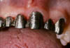
Fig 1: Bulky metal substructure for anterior maxillary porcelain fused to metal bridge.
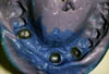
Fig 2: Transfer impression incorporating porcelain fused to metal substructure.
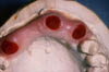
Fig 3: Soft tissue transfer model with GC Pattern Resin abutment replicas.
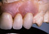
Fig 4: Properly contoured porcelain of anterior maxillary bridgework on the soft tissue transfer model respecting the clinical gingival contour.
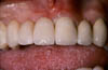
Fig 5: Properly contoured porcelain of anterior maxillary brid-gework intraoral view, respecting the clinical gingival contour.
| 1. | Perel ML. Axial crown contours. J Prosthet Dent 1971; 25:642-9. | |
| 2. | Chiche G, Kokich V, Candill R. Diagnosis and treatment planning of esthetic problems. In: Chiche G, Pinault A. eds. Esthetics of anterior fixed prosthodontics. Chicago: Quintessence Pub. Co. Inc. 1994:39. | |
| 3. | Wilson RD, Maynard G. Intracrevicular restorative dentistry. Int J Periodontics Restorative Dent 1981; 1:35-9. | |
| 4. | Saba S. Anatomically correct soft tis-sue profiles using fixed detachable provisional implant restorations. J Can Dent Assoc 1997; 63:767-70. |