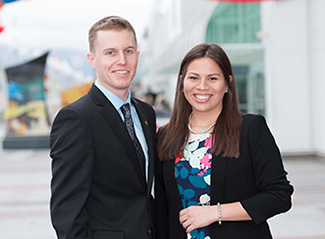
Winners of the 2014 CDA/DENTSPLY Student Clinician Research Program: Tobias Meiszinger, McGill University (2nd Prize); Kellyana Quattrini, University of Saskatchewan (1st Prize)
The 2014 CDA/DENTSPLY Student Clinician Research Program took place in March during the Pacific Dental Conference in Vancouver. The national competition invites dental students from the 10 accredited Canadian dental schools to present research table clinics in front of qualified judges.
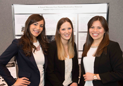
First prize is one expense-paid trip to the American Dental Association (ADA) Annual Session, where the winning table clinic will be presented as part of the ADA’s scientific program. First prize was awarded to a study from the University of Saskatchewan that developed and tested a novel mercury-free metal restorative material. The table clinic was presented by Mrs. Kellyana Quattrini, along with her research collaborators Mrs. Anapaula Campos and Ms. Jenna Schmitt.
“The commitment to research shown by CDA and DENTSPLY is really incredible and vital to the future of our profession,” says Mrs. Quattrini. On behalf of her study collaborators, she adds, “We were very excited to represent our university, and to have the invaluable opportunity to learn from other student clinicians and industry leaders. We look forward to representing the University of Saskatchewan College of Dentistry and CDA/DENTSPLY at the ADA meeting in San Antonio.”
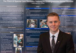
Second prize went to Mr. Tobias Meiszinger of McGill University. Mr. Meiszinger and his research partner, Ms. Gabrielle Lemay, examined special care dentistry training in a Canadian dental school. Mr. Meiszinger received a $1000 cash award for his second-place finish. “What I enjoyed most was the opportunity to use my research findings as a platform to advocate for those with special needs. It was a humbling experience to meet dental professionals who were lifelong advocates for people with special needs, and who recognized the importance of increasing didactic and clinical exposure of dental students to these patients.” says Mr. Meiszigner.
DENTSPLY has sponsored the student clinician research program in Canada since 1971. The program’s purpose is “to stimulate ideas, to improve communication and most of all, to increase student involvement in the advancement of the dental profession.”
The student clinicians participating in the event were also honoured by the Canadian section of the Pierre Fauchard Academy (PFA). The students were presented with a scholarship from the PFA, recognizing their special efforts in the advancement of dental education over and above their academic careers.
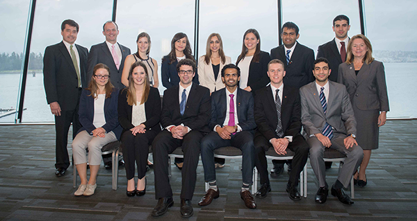
Front row (L. to r.): Sofia Kholmogorova, University of Montreal; Jenna Schmitt, University of Saskatchewan; Richard Miron, Laval University; Gurinder Boparai, University of Manitoba; Tobias Meiszinger, McGill University; Muhammed Al-Nuaimi, University of Alberta.
Back row (L. to r.): David Hancin, vice-president and general manager, DENTSPLY Canada; Dr. Peter Doig, CDA president; Allie Anderson, Dalhousie University; Anapaula Campos, University of Saskatchewan; Natasha, University of Toronto; Kellyana Quattrini, University of Saskatchewan; Joel Keshwah, Schulich School of Medicine and Dentistry; Kambiz Vatandoost, University of British Columbia; Dr. Teresa A. Dolan, vice-president and chief clinical officer, DENTSPLY International.
CDA is pleased to publish the abstracts submitted for the CDA/DENTSPLY 2014 Student Clinician Research Program. To qualify, the study must fall under the “clinical application and techniques” or “basic science and research” categories. Students must identify the purpose of the study, provide background information, outline how the study was conducted and report on the results of the study and its possible significance. The student, selected by his or her own faculty, must be an undergraduate at the time of the presentation, as well as a member of CDA. Ten dental schools participated in this year’s competition.
In some cases, student clinicians collaborated with other students or faculty members to conduct their studies. However, only the names of the official presenters are listed with the abstracts. The abstracts are published in the language of submission.
1st Place: A Novel Mercury-Free Restorative Material
By: Anapaula Campos, Kellyana Quattrini and Jenna Schmitt – University of Saskatchewan
Advisors: Dr. Azita Zerehgar and Dr. Assem Hedayat
Problem: This research aimed to develop and subsequently test a novel restorative material created through compositional modifications of conventional amalgam. Although dental amalgam is commonly used in restorative dentistry, its mercury component is associated with perceived health hazards. Modifications were made to Permite amalgam to eliminate the presumed adverse effects of mercury, impart antimicrobial properties, and reduce the pressure needed to properly condense conventional dental amalgam.
Methods: Conventional regular-set Permite alloy, vinyltriethoxysilane-sialinized titanium dioxide ceramic (VSTDC) nanoparticles (60nm), and a proprietary silver solution were used to create the novel restorative material. Experimentation consisted of completely eliminating the mercury content in Permite amalgam and replacing it with the silver solution and VSTDC nanoparticles. The silver solution and VSTDC nanoparticles were triturated with the Permite alloy.
All components of the mixture were tested in variable amounts until an optimal consistency of the experimental restorative material was obtained. Prepared maxillary premolars were divided into a control group filled with Permite amalgam, and an experimental group filled with the novel material. Both groups were thermocycled, cross-sectioned, and studied through scanning electron microscopy (SEM).
Results: Triturating 600mg Permite alloy, 600mg VSTDC nanoparticles and 3.0ml of silver for 20 seconds yielded the most optimal consistency. SEM images obtained from the control and experimental groups of cross-sectioned teeth revealed superior marginal adaptation of the novel material compared to Permite amalgam; this was achieved by applying reduced condensation pressure in comparison to Permite amalgam.
2nd Place: The Place of Special Care Dentistry Training in Undergraduate Dental Studies
By: Tobias Meiszinger, McGill University
Advisor: Dr. Zovinar der Khatchadourian
Problem: Prior research suggests that North American dental students lack sufficient didactic training about and clinical exposure to patients with special needs (PSN). As a result, graduating students are not adequately prepared to welcome PSN into their private practice.
Methods: A qualitative descriptive case study methodology was used. Data collection included: 1) review of the DMD curriculum from one Canadian dental school; 2) semi-structured interviews of fourth year dental students (n=10) recorded and analysed for themes regarding didactic and clinical educational experiences related to PSN and issues relating to confidence, comfort, and motivation to treat PSN.
Results: Student recall of didactic material was excellent demonstrating great interest in special care dentistry; however, less than 1% of didactic curriculum focuses on PSN.
Supervised clinical exposure (SCE) to PSN helped students challenge pre-existing stereotypes and develop empathy, patience, and communication skills. Learning was compromised due to a lack of previous student exposure to PSN, poor clinical training prior to SCE, and the stressful environment of SCE.
90% of students reported confidence and comfort with communicating with PSN; however, students lacked clinical confidence. Students were aware of their social responsibility to treat PSN yet motivation was hindered for 20% because of stressful SCE experiences.
Didactic material presented in an interactive environment, covering clinical strategies for a wide range of PSN, and strategically scheduled would benefit student learning and ease transition into SCE. Feasible solutions exist to increase student exposure to PSN prior to SCE. Structured student reflection may also consolidate clinical learning experiences.
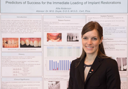
Predictors of Success for the Immediate Loading of Implant Restorations
By: Allie Anderson, Dalhousie University
Advisor: Dr. M. Gorman Doyle
Problem: Implant restorations are becoming increasingly more popular amongst the general public for the replacement of a single missing tooth. Many patients believe that an implant and the restoration can be placed in just one appointment. However, specific criteria need to be satisfied for successful immediate implant restorations.
Methods: A literature review was conducted using PubMed. Various combinations of the following search terms were used: “immediate loading”, “dental implant”, “primary stability”, “implant length”, “loading failure”, “maxillary and mandibular position”, and “insertion torque”. Searches were limited to clinical trials, randomized controlled trials, and systematic reviews written in English.
Results: A comprehensive list of the factors involved in the success of immediate loading of implant restorations was compiled. The factors can be broken down into four main categories: 1) patient, 2) implant, 3) surgical and 4) post-surgical. Each category has several criteria that must be met to ensure long-term success of the implant. Diagnosis using these factors will help clinicians decide if a patient is suitable for immediate loading of an implant restoration.
In conclusion, there are situations where immediate loading of implant restorations is a viable treatment option. The risks and benefits of both immediate loading and conventional delayed loading implant restorations must be discussed with the patient. If the patient has the favorable characteristics, immediate loading of the implant should be considered as a treatment option.
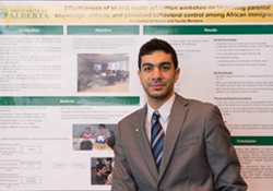
Effectiveness of an Oral Health Education Workshop for African Immigrant Parents.
By: Muhammed Al-Nuaimi, University of Alberta
Advisor: Dr. Maryam Amin
Objectives: To evaluate the effectiveness of a one-session educational workshop on parental knowledge about and attitudes toward oral health of their young children and preventive measures as well as their perceived behavioral control.
Methods: A three-hour oral health education workshop including audio/visual and hands-on components was developed. Participants were parents of young children who had lived in Canada for less than ten years. The workshops were conducted in different community locations by a trained dentist and bilingual community workers who also helped with recruitment, interpretation, and administration of the questionnaires. The impact of the workshop was evaluated using a pre- and a post-intervention questionnaire developed based on theory of planned behavior.
Results: A total of 105 African parents participated in 10 workshops. They were mainly mothers (86.1%) with mean age of 35.03 ± 5.4 years who came to Canada as a refugee (77.1 %) and had three or more children (49.5%). About 70% of the participants had below high school education,
39% were single parent, and 39% had monthly income below $2000. A statistically significant improvement was found in participants’ knowledge about caries and preventive measures, attitudes towards preventive measures, and their perceived behavioural control. Parents’ intention to take their child to a dentist within the next six months also significantly altered after the workshop (p value <0.05).
Conclusions: A hands-on training of newcomer parents in respect to children’s oral health was effective in changing their knowledge, attitudes, and intention to take action in a foreseeable future.
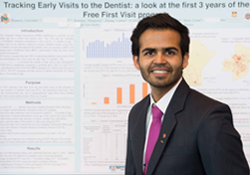
Tracking Early Visits to the Dentist: A Look at the First 3 Years of the Free First Visit Program
By: Gurinder Boparai, University of Manitoba
Advisor: Dr. Robert Schroth
Problem: Early visits to the dentist are important to ensure children have good oral health. The
Free First Visit (FFV) program was launched to provide access to dental screenings for children ≤ 36 months of age. The purpose was to review FFV tracking forms submitted by participating dentists.
Methods: Submitted tracking form data from participating dental offices were reviewed for children ≤36 months of age. These forms collected information on the age of the child at the time of their FFV, their home postal code, if they had any caries, the dentist’s name, and whether they had dental insurance benefits. Descriptive and bivariate analyses were performed. Geo-mapping was completed using ArcGIS.
Results: 8,396 tracking forms were submitted. The mean age at first visit was 24.2 ± 7.8 months. While only 8.5% had a FFV by 12 months of age, 26.7% had a FFV by 18 months of age. Postal code mapping revealed that participation was highest among children in the southern region of the province, including high-needs neighbourhoods within the capital city. Pediatric dentists provided the majority of the FFVs and saw significantly younger children compared to general dentists (23.8 ± 7.8 months vs. 25.2 ± 7.7, p <.001). Results suggest many children participated and received a FFV. However, only a small percentage received visits by 12 months of age.
There is a need to improve the proportion of children receiving first visits by the recommended age and general dentists need to assume a greater role in providing this service.
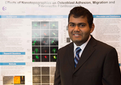
Effects of Nanotopographies on Osteoblast Adhesion, Migration and Fibronectin Fibrillogenesis
By: Joel Keshwah, Schulich School of Medicine and Dentistry
Advisor: Dr. Douglas Hamilton
Problem: The success of dental implants depends on proper balance of bone formation and
resorption. Osteoblasts, which form bone, adhere and sense their environment via focal adhesions. Changes to the substratum topography of implants, is a desirable way to influence osteoblasts, since it persists for the lifespan of the implant. Our study assessed the influence of nanotopographies on osteoblast adhesion, migration and fibrillogenesis.
Methods: Rat calvarial osteoblasts were seated on glass slides, with nanotopographies etched into the surface (lines, dots). Immunocytochemistry was used to assess adhesion site anchoring proteins vinculin, tensin and extracellular matrix (ECM) molecule fibronectin. Cell migration was analyzed using time-lapse video (TLV).
Results: Focal contacts formed at the peaks of topographies in cells on dot surfaces, while focal contacts formed parallel to the long axis of the topography on grooved surfaces. Cells on smooth surfaces formed large adhesion plaques with random orientation. Fibrillar adhesions showed similar, but less pronounced patterning as focal contacts. TLV showed cells on line surfaces migrated in the direction of the topography while cells on other surfaces migrated randomly. No significant difference was seen in migration velocity or distance travelled, but cells on lines and dots tend to travel faster and farther. After 72 hours, cells on grooved surfaces developed fibronectin fibrils parallel with the topography, while fibronectin on dot and smooth surfaces had a random orientation. In conclusion, substratum nanotopographies can manipulate osteoblast physiology and by understanding its impact, novel implants that facilitate osseointegration through effects on bone remodeling can be designed.
Integrity of the Junctional Epithelium: Structural and Functional Studies of Novel Inner Basement Membrane Components
By: Sofia Kholmogorova, University of Montreal
Advisor: Dr. Annie St-Georges
Functional genomic screening of the rat enamel organ has led to the identification of Amelotin (AMTN), Odontogenic ameloblast-associated (ODAM) and SCPP-Proline- Glutamine-rich 1 (SCPPPQ1) as novel secretory products of maturation stage ameloblasts. These proteins are also produced by the junctional epithelium (JE) and localize to the basal lamina of the epithelial attachment where they seal off the periodontium from the aggressive oral environment. Understanding how the JE binds to the tooth is therefore of high clinical relevance. We have exploited atomic force microscopy (AFM) to obtain structural information on recombinant rat ODAM, AMTN and SCPPPQ1, to visualize their potential interactions. Each protein resulted in a distinct pattern of distribution on an atomically neutral surface. When they were added sequentially to the substrate, change in the resulting pattern suggested protein interactions. Images of AMTN layered onto SCPPPQ1 revealed intriguing 100 nm pores in a homogenous background matrix. Interestingly, the addition of ODAM over the layered SCPPPQ1 and AMTN resulted in a reduction of the pore size. These data suggest that AMTN, ODAM and SCPPPQ1 are capable of self-assembly and that protein-protein interactions occur among them. These analyses represent a first step towards exploring the molecular arrangement and interactions of proteins in the JE basal lamina and how these result in a unique adhesive relationship with the tooth. The application of one or all of these proteins may potentially represent a novel biomimetic therapeutic strategy to maintain and restore JE integrity before periodontal disease causes the inevitable loss of the supporting structures of the tooth.
Osteoinductive Potential of Commonly Employed Bone Grafts – The Development of Vivoss
By: Richard Miron, Laval University
Advisor: Dr. Fatiha Chandad
Since the original description of osteoinduction in the early 20th century, the development of innovative biomaterials with osteoinductive potential has emerged. Over the last 50 years however, our ability to accurately describe biological phenomenons has been drastically improved by advancements in cell and molecular biology. The aim of the present study was to compare the osteoinductive potential of bone grafts by comparing autogenous bone, demineralized freeze-dried bone allografts (DFDBA), a commonly utilised xenograft (BioOss) and a newly developed biphasic calcium phosphate. Grafts were compared in vitro for their ability to stimulate mesenchymal cell migration, proliferation and differentiation as assessed by alkaline phosphatise (ALP) activity and real-time PCR for bone-markers including Runx2, ALP, collagenI, and osteocalcin. Furthermore, bone grafts were implanted in the calf muscle of beagle dogs to determine their potential to form ectopic bone in vivo. The results from the present study demonstrate that while xenografts were not osteoinductive and autogenous bone grafts were resorbed quickly in vivo, major differences in the bone inducing capabilities between DFDBA and synthetic biphasic calcium phosphate alloplasts were observed. The modifications in nanotopography and chemical composition of the new biphasic calcium phosphate bone graft significantly improved osteoblast differentiation and promoted ectopic bone formation. Although the future field of osteoinductive biomaterials faces many challenges in order to meet the incoming demand for bone grafting procedures worldwide, the results from the present study provide evidence that modifications to synthetic bone grafts no longer only serve as a 3-dimensional scaffold but are also able to spontaneously induce bone formation.
Novel Minimally-Invasive Means to Survey Bacterial Biomarkers Under Chronic Periodontitis
By: Natasha, University of Toronto
Advisor: Dr. Dilani Senadheera
Problem: Periodontitis, a significant oral health burden, is a consequence of microbial dysbiosis that elicits an inflammatory response destroying tooth-supporting structures. Of several varieties, chronic periodontitis (CP) remains the leading cause of tooth exfoliation worldwide. Conventionally, treatment outcomes against periodontitis are monitored via culturing/immunostaining of odontopathogens in subgingival (SubG) plaque. Hypotheses: In lieu of SubG sampling, selected bacterial biomarkers associated with CP are quantifiable using supragingival (SupG) or tongue plaque by determining bacterial relative abundance (RA) with real-time PCR (RtPCR).
Methods: DNA was extracted from 72 plaque samples (SupG, SubG and tongue sites) from 11-healthy and 13-CP individuals. RtPCR was used to quantify bacterial load and RAs of Tannerella forsythia (Tf), Porphyromonas gingivalis (Pg), Rothia dentocariosa (Rd), Filifactor alocis (Fa) with 16S rRNA gene- specific primers. Statistical analysis: Two-way-ANOVA/ Tukey's post-hoc test.
Results: Bacterial load was unaffected by disease status. Pg and Tf represent the most abundant taxa under CP, with significantly increased Pg levels in all sampling sites. Rd numbers were increased under health. Tf and Fa levels were drastically elevated in SubG and SupG sites, although Fa increase was not statistically significant.
Conclusions: SupG and tongue sites harbor significantly altered levels of selected bacterial biomarkers depending on periodontal health that are quantifiable with RtPCR. Presently, we are examining RAs of >15 putative periodontal pathogens in different oral sites. Here, we report a novel and convenient method to sample and quantify CP-associated taxa that provides critical groundwork to develop a rapid and minimally-invasive strategy to monitor treatment modalities against CP.
Differential Regulation of Keratincyte Function by Skin and Gingival Fibroblasts
By: Kambiz Vatandoost, University of British Columbia
Advisor: Dr. Ravindra M. Shah
Objectives: Human gingiva has fast wound healing that involves rapid re-epithelialization and results to minimal scaring compared to skin wounds. Mechanisms for this difference are poorly understood but may depend on distinct phenotype of skin and gingival fibroblasts. Wound healing is regulated by cross-talk between epithelium and connective tissue cells. Therefore, we hypothesized that gingival and skin fibroblasts secrete soluble mediators that regulate gene expression and affect function of epithelial keratinocytes differently. To this end, we compared gene expression in human keratinocytes treated with conditioned medium (CM) from human gingival and skin fibroblasts propagated in in vivo-like three-dimensional (3D) cultures.
Methods: Fibroblasts from three parallel cell lines isolated from human skin (breast) and gingiva were cultured in an in vivo-like 3D culture, CM was collected and used to treat skin keratinocytes (HaCaT). Expression of key genes involved in wound healing and inflammation was analyzed by real-time RT-PCR.
Results: In general, CM from both gingival and skin fibroblasts regulated the expression of integrins, certain ECM proteins, cytokines, MMPs and cell proliferation, angiogenesis and cell communication-associated genes in keratinocytes. Expression of a set of genes involved in cell-ECM interactions, inflammation and angiogenesis were differently regulated by gingival and skin fibroblast CM in the keratinocytes.
Conclusions: Distinct composition of molecules secreted by skin and gingival fibroblasts regulate keratinocyte’s phenotype differently. This may contribute to the different wound-healing outcomes in skin and gingiva.
**
Photos by Teckles Photos Inc.