|
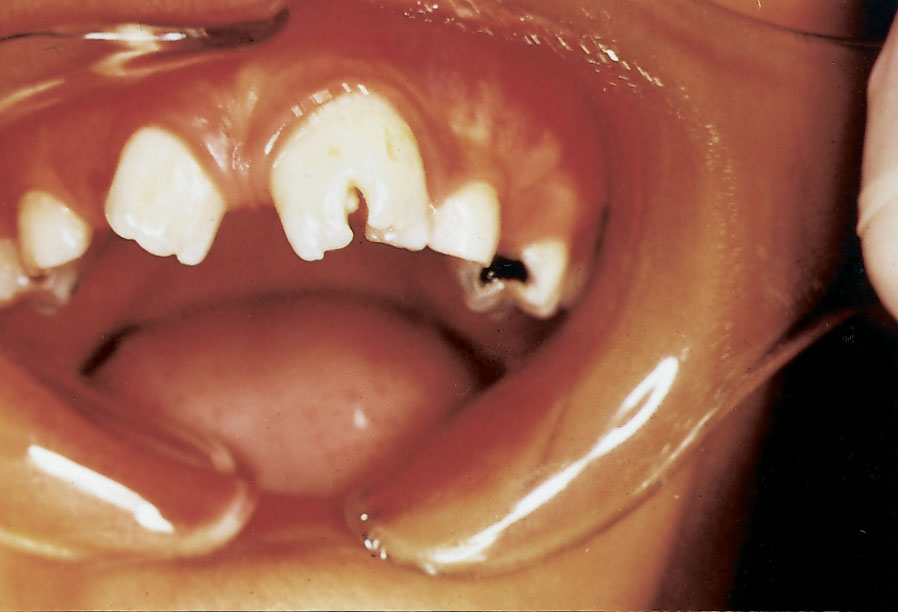
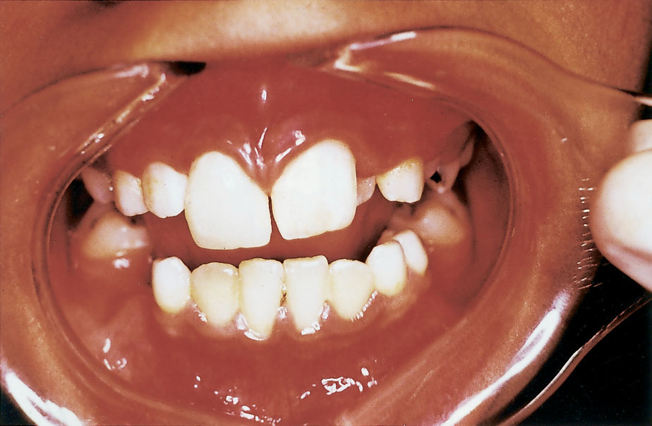
| Figure 1: Frontal view on initial presentation showing large, bifid and malpositioned geminated maxillary left central incisor associated with wide midline diastema. |
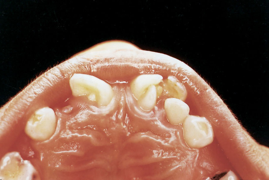 |
| Figure 2: Palatal view demonstrating talon cusp on the geminated left central incisor projecting from the cingulum and extending almost to the incisal edge. The right central incisor exhibits exaggerated cingulum. |
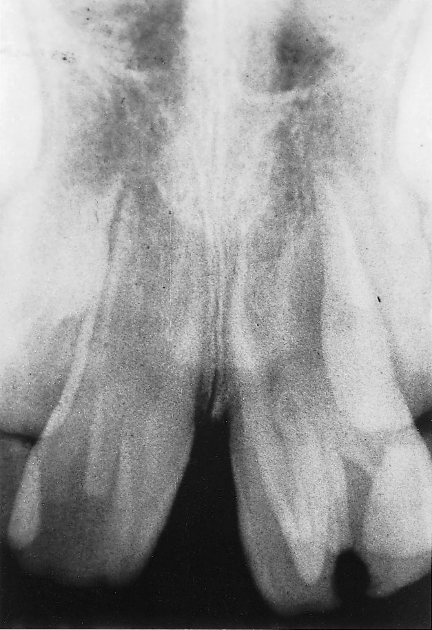 |
| Figure 3: Periapical radiograph showing talon cusp outlined by two white lines representing the enamel, with pulp horn extending to the middle of the cusp. Note the enlarged pulp chamber and the single root of the affected tooth. |
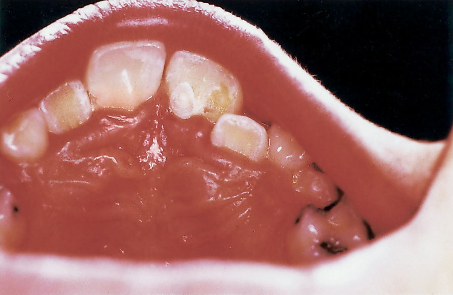 |
| Figure 4: The talon cusp was reduced almost completely after 3 visits at 6- to 8-week intervals without exposing the pulp. |
 |
| Figure 5: Appearance of the maxillary incisors after conservative and orthodontic treatment. |