|
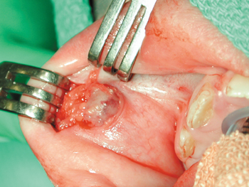
| Figure 1: Exposure of a rapidly growing intraoral lesion in a 9-year-old girl. |
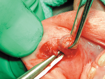 |
| Figure 2: Removal of the lesion. |
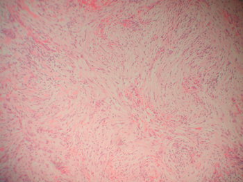 |
| Figure 3: Histopathological examination, using haematoxylin and eosin (H & E) stain, reveals the storiform pattern. (Original magnication × 100.) |
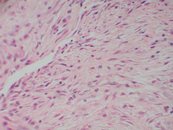 |
| Figure 4: Histopathological examination using H & E stain, in the centre of the nodule, showing spindle cells. (Original magnigcation × 400.) |
