|
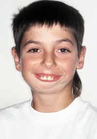
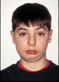
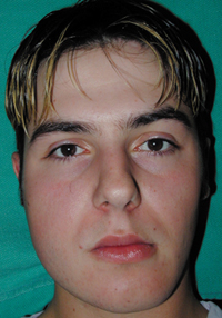
| Figure 1a: Pretreatment frontal view. |
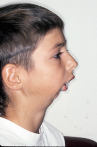 |
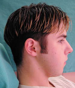
| Figure 1b: Pretreatment - lateral view. |
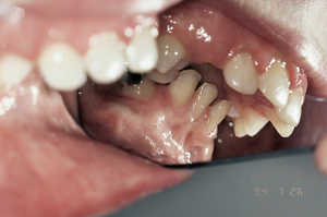 |
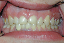
| Figure 1c: Pretreatment — occlusal view. |
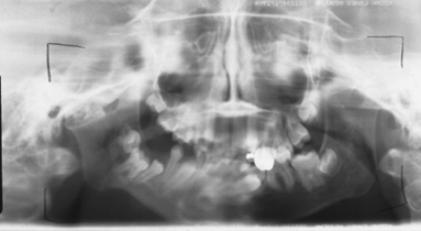 |
| Figure 2a: Pretreatment — panoramic radiograph. |
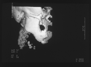 |
| Figure 2b: Pretreatment — 3-dimensional computed tomogram. |
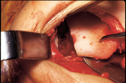 |
| Figure 3: Gap arthroplasty. |
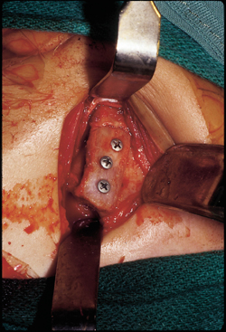 |
| Figure 4: Costochondral graft secured to mandibular ramus with 3 bicortical screws. |
 |
| Figure 5: Post-treatment, 2 years — frontal view. |
 |
| Figure 6a: Post-treatment, 8 years — frontal view. |
 |
| Figure 6b: Post-treatment, 8 years — lateral view. |
 |
| Figure 6c: Post-treatment, 8 years — occlusal view. |
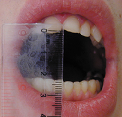 |
| Figure 6d: Post-treatment, 8 years — maximum opening. |