Fig.1

Russell1.jpg (62154 bytes)
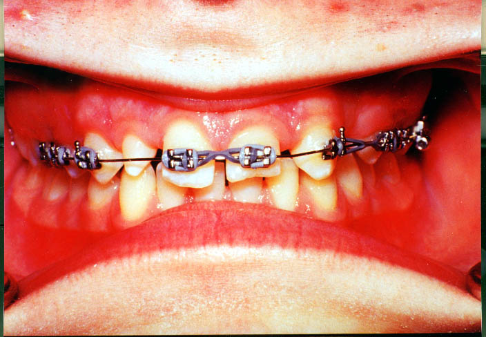
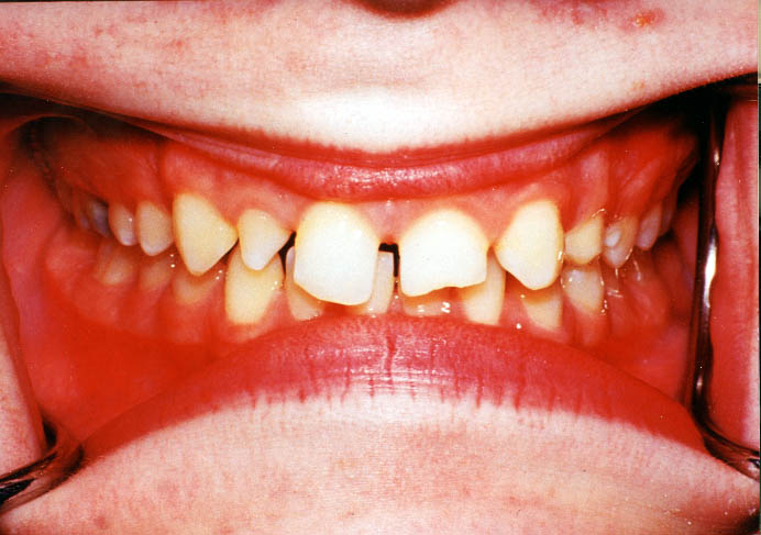
Figure 1: Pretreatment intraoral photograph of a congenitally missing lateral incisor and canine that has drifted mesially. Preprosthetic orthodontic treatment is necessary to align the crowns and roots of the central incisor and canine before implant restoration.


Fig.2

Russell2.jpg (74123 bytes)
Figure 2: Mid-treatment intraoral view showing sufficient space between crowns for placement of a traditional fixed or removable prosthesis.


Fig.3

Russell3.jpg (36707 bytes)
Figure 3: Mid-treatment panoramic radiograph revealing insufficient space between the roots of the teeth for placement of the implant, although there is sufficient space between the crowns.


Fig.4

Russell4.jpg (72382 bytes)
Figure 4: The final orthodontic positions showing sufficient space between crowns for the implant and sufficient space to build up the contra-lateral peg-lateral.


Fig.5

Russell5.jpg (37578 bytes)
Figure 5: Sufficient space between the roots has been created with additional orthodontic treatment by moving the central incisor and canine roots further apart to allow placement of the implant fixture.


Fig. 6

Russell6.jpg (85132 bytes)
Figure 6: Vacuum-form retainer in place following orthodontic treatment. A denture tooth has been placed in the retainer to maintain the space for the implant restoration