|
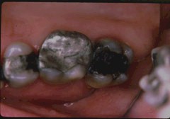
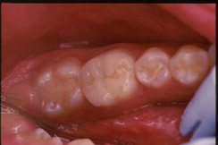
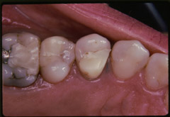
| Figure 1: Judging by the extent of corrosion, amalgam restorations on maxillary second premolar and second molar teeth were probably fabricated from a conventional amalgam alloy. The one on the first molar shows much less corrosion, indicating that it could have been made from a high-copper amalgam alloy. |
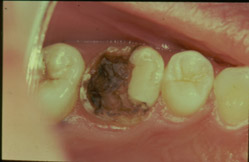 |
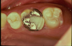
| Figure 2: Left maxillary first molar of a 12-year-old girl with lost amalgam restoration and evidence of recurrent caries. |
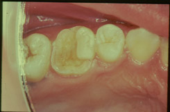 |
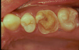
| Figure 3: Cavity preparation for an adhesive resin composite onlay restoration. |
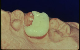 |
| Figure 4: The dentist-fabricated onlay restoration seated on the stone model. |
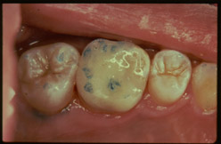 |
| Figure 5: Occlusal adjustment was done after rubber dam removal. |
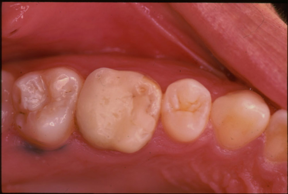 |
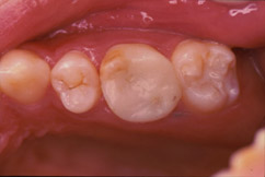
| Figure 6: View of the cemented onlay restoration two years after placement. |
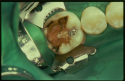 |
| Figure 7: This massive carious lesion on lower first molar tooth occurred underneath an existing amalgam restoration. |
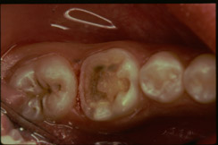 |
| Figure 8: After three months of pulp-capping, the tooth was prepared to receive an onlay ceramic restoration. |
 |
| Figure 9: View of the cemented IPS Impress onlay restoration nine months after placement. |
 |
| Figure 10: This maxillary first molar presented with a combined amalgam/resin composite restoration with evidence of breakdown. |
 |
| Figure 11: Completed cavity preparation for an adhesive resin composite restoration. |
 |
| Figure 12: Completed direct resin composite restoration eight months after placement. |
 |
| Figure 13: Large resin composite restorations on maxillary premolars eight years after placement. These were made using the material P-50 (3M). |