Orthodontic Management of Missing Teeth
• David B. Kennedy, BDS, LDS RCS (Eng), MSD, FRCD(C) •
© J Can Dent Assoc 1999; 65:548-50
[Diagnosis and Treatment Planning Considerations |Cost |References]
Approximately 2% to 10% of the population exhibit missing teeth. Excluding third
molars, the most commonly missing teeth are maxillary lateral incisors and second
premolars. Patients who exhibit congenital absence of teeth also experience increased
ectopic dental eruption and other dental anomalies (Fig. 1).1
Specifically, patients with missing lateral incisors frequently have contralateral lateral
incisors peg-shaped or smaller than the normal mesial distal width. Patients with
congenitally absent maxillary lateral incisors often exhibit palatal ectopic eruption of
the adjacent maxillary permanent canines.1,2 Permanent canines adjacent to
absent lateral incisors also erupt mesially. In cases of unilateral absence of a maxillary
lateral incisor, the midline is often deviated toward that side (Fig. 2).
 |
 |
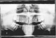
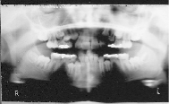
| Figure 1: Panoramic x-ray shows ectopic
canine, absent lower right second premolar, ankylosed primary second molar, ectopic lower
left second premolar and absent third molars. |
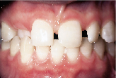
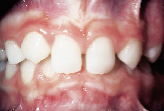
Figure 2: Pre-orthodontic anterior
view showing missing right lateral incisor, small left lateral incisor and maxillary
midline deflected to right side with ectopic palatal upper canine. |
Second primary molars without underlying permanent successors show increased frequency
of ankylosis and submergence.1 The earlier that ankylosis exists, the more
severe are the potential consequences. Progressive infraocclusion can result in
compromised alveolar bone height, tipping of adjacent teeth and destruction of bone during
the extraction. Because infraocclusion increases as vertical bone development occurs
commensurate with facial growth, the earlier the infraocclusion occurs, the more necessary
it is to have the primary molar extracted to preserve vertical alveolar bone. The larger
mesiodistal width of a retained second primary molar compared to the smaller width of the
absent second premolar can compromise molar occlusion.
 [ Top ]
Diagnosis and Treatment Planning Considerations
General Principles
The clinician should always ask two questions when faced with a patient who has
congenital absence of permanent teeth.
1. What would you (the clinician) do if the missing tooth were indeed present?
2. Can this malocclusion be treated satisfactorily with extractions or not?
If the malocclusion can be satisfactorily treated with extraction, then the appropriate
removal of the primary tooth (which has no permanent successor) can permit orthodontic
space closure; thus, the congenital absence of the tooth has less long-term consequence.
By contrast, if extractions are contraindicated, then consideration has to be given to the
long-term prosthetic management of the area where the permanent tooth is absent. Such
management includes the management of any retained primary teeth (such as second primary
molars where second premolars are absent).
The patient needs to be thoroughly diagnosed using a planes-of-space concept.3
Once a problem list has been established, then treatment objectives can be developed to
meet the patient’s needs. Based on these treatment goals, appropriate treatment
alternatives can be investigated.3 Often, a diagnostic wax setup study model is
needed to show the referring dentist, the patient and the parent the estimated final
occlusion and the possible position of artificial tooth replacement. This diagnostic wax
setup can determine anchorage requirements and help formulate a mechanical treatment plan.
Furthermore, it can determine appropriate pontic widths, potential tooth-size
discrepancies and the need for post-treatment bonding or interproximal tooth reduction. It
also allows the restorative dentist the opportunity for input in the treatment planning
stage (Figs. 2, 3 and 4). Obviously, this treatment planning exercise can be
done only with comprehensive orthodontic records.
|
 |
 |
| Figure 2: Pre-orthodontic
anterior view showing missing right lateral incisor, small left lateral incisor and
maxillary midline deflected to right side with ectopic palatal upper canine. |
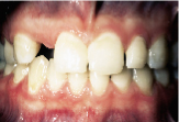
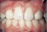
Figure 3: Post-orthodontic view of
patient seen in Figure 2. Midlines centred. Appropriate space opened up for prosthetic
management of missing lateral incisor and small left lateral incisor. |
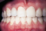
Figure 4: Post-restorative view of
patient seen in Figures 2 and 3. Implant and crown replaces missing right lateral incisor.
Porcelain veneer on left lateral incisor. Cosmetic bonding reshapes central incisors. |
[ Top ]
The Missing Maxillary Lateral Incisor
Esthetic requirements usually dictate space opening and subsequent
post-orthodontic artificial replacement of the missing lateral incisor or incisors. The
two instances where extraction and space closure (of the missing lateral incisors) would
be appropriate are:
1. in patients with sufficient crowding to warrant an extraction treatment plan and who
have congenital absence of one or both lateral incisors (Fig. 5) and
2. Class II malocclusions with an acceptable facial profile that can be satisfactorily
treated with either a single upper arch extraction plan or with upper and lower arch
extractions.
 |
 |
| Figure 5: Absent maxillary
lateral incisors, deep overbite and overretained right primary canine. Pretreatment view. |
Figure 6: Post-treatment view.
Absent maxillary lateral incisor spaces closed due to crowding. Canines reshaped and
bonded. |
There are at least six major disadvantages to closing missing maxillary lateral incisor
spaces:
1. Pointed maxillary canines require post-orthodontic grinding or cosmetic bonding to
simulate an incisor (Figs. 5 and 6).
2. Maxillary canines are usually darker than lateral incisors; veneering may be
necessary.
3. Maxillary canines are wider than adjacent absent lateral incisors, creating an
esthetic mismatch and an anterior tooth-size discrepancy. The six upper anterior teeth
(first premolar, canine and central incisor) are relatively too wide for the corresponding
lower six anterior teeth (canine, lateral and central incisor). This discrepancy can cause
an increased overjet unless interproximal reduction is contemplated.
4. In canine substitution cases, the first premolar serves as a canine; the lingual
cusp often needs to be reduced for esthetic or functional reasons.
5. Because the labiolingual thickness of the upper canine is greater than the
corresponding missing lateral incisor, selective palatal reduction of the canine is often
needed.
6. The final occlusion demonstrates group function rather than canine guidance.
[ Top ]
Cost
Most patients with congenitally absent teeth require comprehensive orthodontic
treatment. If the decision is made during treatment planning for spaces to be closed, then
the patient needs no post-orthodontic restorative dentistry. By contrast, when pontic
spaces are to be subsequently restored, the patient incurs restorative costs for
bridgework or implants, possible periodontal costs for crown lengthening and possible
endodontic costs in instances of repeated tooth preparation (which results in loss of
tooth vitality). Furthermore, replacement of the restoration may be required two or three
times during the patient’s lifetime.4
Implants can be used in non-growing patients. They require a two-stage surgical
procedure and, at present, are not covered by insurance. Additionally, patients who are
candidates for implants may require bone augmentation, because patients without permanent
teeth may not develop the alveolar bone that accompanies eruption.
[ Top ]
Post-Orthodontic Restorative Choices
Immediately post-debanding, removable retainers with denture teeth are used for
full-time wear. Most adolescents and adults prefer fixed prosthesis for replacement of
missing teeth. In most instances, implants supporting crowns are the preferred restorative
choice, because they save tooth destruction of virgin abutment teeth (Fig. 4).
Implants can be placed only after all vertical alveolar growth has ceased. In a girl,
growth cessation may occur by age 15, but boys and young men may not complete growth until
their early 20s. Placing an implant in a growing individual invites submergence, as the
implant behaves like an ankylosed tooth; the resulting clinical crown length and emergence
profile of the restoration are highly undesirable. Because teenagers usually do not want
to wear removable retainers from the completion of orthodontics until they may be ready
for implants, minimal tooth-reduction Maryland bridges can serve as useful interim
restorations.
[ Top ]
The Extraction/Non-Extraction Dilemma for Missing Premolars
Despite rhetoric to the contrary, scientific evidence suggests that appropriate
extraction followed by space closure affects the facial profile negligibly.5
Early extraction of second primary molars in instances of congenital absence of underlying
second premolars can often result in mesial drifting of the posterior dentition, affording
the opportunity to close spaces.6 Therefore, aggressive early modified serial
extraction can eliminate the need for major restorative dentistry.
When crowding is severe, space maintenance is needed to conserve anchorage while
canines and first premolars drift distally, or extractions can be delayed until the
permanent dentition (Figs. 7 and 8). In Class II cases, anchorage
requirements dictate extraction timing and mechanical space closure management.
 |
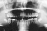 |
| Figure 7: Pre-treatment panoramic x-ray
shows missing lower second premolars and developing canine crowding. Case managed by
extraction of upper first premolars and lower second primary molars. |
Figure 8: Post-orthodontic
panoramic x-ray shows absent lower second premolar spaces closed and extraction of upper
first premolars. |
By contrast, later extraction of premolars can have a more negative effect on incisor
position7 and therefore can result in flattening of the facial profile if there
is excessive incisor retraction. For these reasons, it is imperative that patients be
assessed orthodontically as early as possible so that all treatment options are available.
During treatment planning, the following factors increase the likelihood of extraction:
1. a large degree of crowding,
2. midline discrepancy,
3. anteroposterior molar discrepancy between right and left sides,
4. procumbency of the incisors on the underlying alveolar structures (double-dental
protrusion),
5. full facial profile requiring reduction in lip support,
6. increased vertical dimension of the lower facial height, and
7. a shallow overbite or anterior openbite.
[ Top ]
Dental Health Considerations
There is no evidence that temporomandibular joint health is compromised by
either orthodontics or extractions.8 Therefore, this should not be a factor in
decision making. The periodontal health of patients with missing lateral incisors treated
by space opening versus space closing shows some differences in response. While the
esthetics were markedly improved in instances where spaces had been opened, long-term
periodontal health was compromised secondary to restorative care.9 Translating
this information to patients with missing premolars, space closure is the treatment of
choice in the posterior region wherever possible
[ Top ]
Coordinated Treatment Planning
Emphasis has been placed on the need to involve the patient and the restorative
dentist in the treatment plan. Because patients with congenitally missing teeth have other
dental anomalies associated, it is imperative that they be supervised closely from an
early age. The patient is afforded more treatment opportunities if seen at the age of
early mixed dentition rather than only at the age of early permanent dentition.
During the final stages of orthodontic care, referral to the diagnostic wax setup helps
check the targeted final tooth position and pontic size. Referral to the restorative
dentist before orthodontic appliance removal allows the restorative dentist input into
final tooth position.
[ Top ]
Acknowledgment: The restorative care shown in Figure 4 was provided by Dr.
David Bridger of Vancouver, B.C.
Dr. Kennedy maintains a private practice in Vancouver B.C. He specializes in
orthodontics and pediatric dentistry.
Reprint requests to: Dr. David B. Kennedy, 200-650 West 41st Ave., Vancouver, BC
V5Z 2M9
The author has no declared financial interest in any company manufacturing the types of
products mentioned in this article.
[ Top ]
References
1. Baccetti T. A controlled study of associated dental anomalies. Angle
Orthod 1998; 68:267-74.
2. Peck S, Peck L, Kataja M. Prevalence of tooth agenesis and peg-shaped maxillary
lateral incisor associated with palatally displaced canine (PDC) anomaly. Amer J Orthod
Dentofacial Orthop 1996; 110:441-3.
3. Proffit WR, Ackerman JL, Fields HW. Diagnosis and treatment planning in
orthodontics. In: Proffit WR, Fields HW, editors. Contemporary Orthodontics. 2nd
ed. St-Louis: CV Mosby; 1993. p. 139-225.
4. Creugars N. Two-thirds of fixed partial prosthesis (bridges) last 15 years. Evidence-based
Dentistry 1998; 1:19.
5. Luppanapornlarp S, Johnston LE Jr. The effects of premolar extraction: a long-term
comparison of outcomes in “clear-cut” extraction and non-extraction Class II
patients. Angle Orthod 1993; 63:257-72.
6. Joondeph DR, and McNeill RW. Congenitally absent second premolars: an interceptive
approach. Amer J Orthod 1971; 59:50-66.
7. Papandreas SG, Buschang PH, Alexander RG, Kennedy DB, Koyama I. Physiologic drift of
the mandibular dentition following first premolar extractions. Angle Orthod 1993;
63:127-36.
8. Artun J, Hollender LG, Truelove EL. Relationship between orthodontic treatment,
condylar position, and internal derangement in the temporomandibular joint. Amer J
Orthod Dentofacial Orthop 1992; 101:48-53.
9. Nordquist GG, McNeill RW. Othodontic vs. restorative treatment of the congenitally
absent lateral incisor — long-term periodontal and occlusal evaluation. J
Periodontol 1975; 46:139-43.
[ Top ]