

![]()
![]()
Predictably Restoring Endodontically Treated Teeth
Alex McLean, DMD, B.Sc. (Eng.)
ABSTRACT
Endodontically treated teeth can be restored with a wide range of techniques of varying complexity. This paper presents a straightforward technique for the restoration of endodontically treated teeth that meet certain standards. Criteria are provided for the utilization of crowns, composite resins, cast gold post cores, amalgam or composite build-ups, and passive, parallel, small diameter stainless steel posts to restore these teeth. Consideration is given to ferrule design and its importance in achieving success.
MeSH Key Words:dental restoration, permanent/methods; patient care planning; post and core technique; tooth, non-vital/therapy.
© J Can Dent Assoc 1998; 64:782-7
This article has been peer reviewed.
[Choices in Build-up Materials | Cast Gold | Amalgam | Composite | Glass Ionomer Ranking of Build-up Materials | How to Retain the Build-up | The Choice of Final Restoration | Ferrule Design | When to Place a Post [Passive Metal Posts | Active Posts | Carbon Fibre Posts | Cast or Preformed Post | Post Head Design [Post Cements | Post Hole Preparation | Discussion | Summary]
A preceding paper1 provided criteria to help identify a predictably restorable endodontically treated tooth. The criteria for an endodontically treated tooth requiring a post is that the minimum length of remaining solid tooth equal the sum of the biologic width (2.5 mm), the ferrule length (2 mm), the apical seal (4 mm) and the post length, or 8.5 mm + post length (Fig. 1). For teeth not requiring a post, the requirements are for biologic width + ferrule length, or 4.5 mm of supra-bony solid tooth. Solid tooth refers to dentin that is a minimum of 1 mm thick after preparation. In addition, consideration of the functional loads on a tooth is essential. Single abutments supporting precision attachment removable partial dentures (RPDs), distal extension RPDs or cantilever fixed partial dentures (FPDs) that are endodontically treated or likely to become so in the future should be avoided. Careful assessment of the occlusal demands and other loads such as FPDs or RPDs must be made prior to restoration.
Fig. 1 Minimum dimensions for a predicably restorable tooth.

Any endodontically treated tooth will require a build-up, which may be as simple as closing the access and pulp chamber on an intact anterior. The optimal build-up material will have adequate strength, be biocompatible, exhibit a high level of resistance to bacterial leakage, and be insoluble and dimensionally stable in the presence of oral fluids. Bullard and others2 showed that a coefficient of thermal expansion close to that of the tooth reduces leakage, which is important. This paper considers the four materials commonly utilized today for build-ups: cast gold, amalgam, composite, and glass ionomer.
Cast gold offers strength. Its resistance to leakage is derived from the luting agent. It doesn't absorb water and has a coefficient of thermal expansion (CTE) very close to that of dentin. Typical CTE for gold3 is 14 x 10-6; the CTE of dentin4 is 10.6 x 10⁻⁶ . Cast gold build-ups require a post for retention and a substantial degree of coronal destruction to be used. Where applicable, this is the build-up material of choice.
Amalgam offers strength. Its coefficient of thermal expansion is almost double that of dentin (about 22 x 10-6, versus 10.6 x 10⁻⁶), and it is relatively stable in the presence of water. It offers a high-level resistance to leakage once it has been in place for a period of time due to the sealing effects of its corrosion products. Initial leakage has been shown to be significantly lower with dispersed phase alloys than with spherical5 alloys. Bonding of amalgams is an option, and has the potential to strengthen the tooth and reduce leakage. Christensen6 recommends that bonding of amalgam restorations be routine; however, with root-treated teeth, sensitivity is not an issue and the additional strength obtained is apparently transient. Santos and Meiers7 found no significant strengthening of teeth with amalgam bond after thermocycling, and Bonilla and White8 found short-term increases in strength that disappeared after 500-day storage or load cycling. As apparently the bond of amalgam to dentin will ultimately degrade, there is concern about increased leakage after bond failure. The bonded surface of the amalgam may be more corrosion resistant than an unbonded amalgam, leading to the risk of increased leakage on a long-term basis.9 Given the short-term nature of the bond, the additional time and cost to bond, and unanswered questions as to the effects on long-term leakage, amalgam bonding is contraindicated for build-ups. On posterior teeth with enough pulp chamber depth to obviate the need for a post (2 to 4 mm), amalgam is the material of choice. Where a post is required to retain the build-up, amalgam is cheaper and faster than a cast gold core and often less destructive of tooth structure.
Composite apparently offers adequate strength clinically, its ultimate strength being somewhat lower than that of amalgam. Its resistance to leakage is almost totally dependent on the luting agent, and the ability of dentin bonding agents to prevent leakage over the long term is unproven. Burrow and others10 showed a degradation of dentin bond strength in vitro over three years almost to the level of an unbonded restoration. If this is typical clinically, then the long-term ability of dentin adhesives to reduce leakage cannot be relied on, which means composite build-ups must rely on mechanical retention as do amalgam build-ups. The coefficient of thermal expansion for most modern self-polymerising composite build-up materials is significantly higher than that of tooth; examples include Ticore11 by EDS at 34 x 10-6, and BisCore12 by Bisco at 25 x 10-6. In addition, composites show significant setting shrinkage. Sakaguchi and others13 showed 0.2% post gel contraction. This shrinkage results in stresses on the bonding systems that may contribute to long-term bond failure. On anterior teeth where a crown is not required and enamel margins offer the promise of long-term resistance to leakage, composite is an excellent choice. With the poor long-term prognosis for dentin bonding agents and the corresponding risk of leakage, relying on composite build-up materials for leakage control seems risky and unpredictable. On posterior teeth where composite is used as a build-up material, maintaining at least 2 mm between crown margins and the build-up should reduce leakage. The absorption of water with composites is a potential concern due to the generation of internal stresses, but it is difficult to assess the clinical significance of this concern. In addition, it has been shown that the mechanical properties of composites degrade with thermocycling and exposure to water.14 Kovarik and others15 showed significantly shorter fatigue life for composite build-up supported crowns than for those with amalgam build-ups. While composites are fast and convenient to use, in most instances they are inferior to amalgam and gold.
Glass ionomer filling materials offer a low level of leakage,16,2 a relatively weak dentin bond and a low level of mechanical strength (Table I). They offer the appeal of fluoride release to reduce decay potential, but there is minimal evidence that this has any clinical significance. Kovarik and others15 fatigue-tested crowns with amalgam, composite and glass ionomer cores, and found that amalgam was significantly stronger than composite and that glass ionomer had inadequate strength as a core build-up. Because of its weak mechanical properties, glass ionomer has little to offer as a build-up material and should be reserved for limited applications such as blocking out minor undercuts.
| Table 1 | |||
Tensile Strength MPa |
Young’s Modulus GPa |
Coefficient of Thermal Expansion x10-6 °C |
|
Cast Gold Type IV3 |
701 to 786 |
69 to 110 |
14 |
Amalgam |
65.717 |
38 to 6018 |
222 |
Composite Biscore12/Ticore11 |
55/35 |
13/18.5 |
25/35 |
Glass Ionomer |
12.419 |
9.419 |
142 |
Dentin |
59.620 |
10.221 |
10.64 |
When a post is required and there is sufficient coronal destruction to allow cast gold without significant additional loss of tooth structure, cast gold is best. In situations where a build-up can be retained without a post, or where a post is needed but the placement of a cast gold core build-up would require significant removal of additional tooth structure, amalgam would be the material of choice. In teeth where simple closure of the access preparation is all that is required and enamel margins offer the potential for long-term resistance to leakage, composite resin is the material of choice. Glass ionomer is not suitable for build-ups.
With the use of cast gold, a post is mandatory for retention. With amalgam or composite, the retentive options are pins, posts and mechanical undercuts such as offered by a pulp chamber. The use of pins to retain build-ups carries the risk of microfracture and introduces stress into the adjacent dentin. Teeth that require pins or posts to retain build-ups have already suffered significant coronal destruction. Caputo21 recommends a minimum of 1 mm of solid dentin to surround a pin, yet this thickness is rarely available to retain build-ups on root-treated teeth. The undercuts in pulp chambers can be used to retain build-ups; Nayyar and others22 and Kane and others23 have shown that pulp chamber depths of 2 to 4 mm offer adequate retention for an amalgam build-up without pins or posts. The use of slots or dovetails cut into dentin to retain the amalgam has appeal but requires the destruction of more tooth structure. There is also a risk that these retentive devices will be removed or weakened during crown preparation.
Posts offer the ability to retain the build-up and, if carefully placed, remove little tooth structure. They are also removable for orthograde endodontic retreatment.
The Choice of Final Restoration
Crowns placed on anterior teeth do not make teeth inherently stronger.24,25 In the absence of significant coronal destruction, a tooth is better restored simply by closing the access with a composite resin. Once distal to the cuspid, placement of a cast restoration that shoes the cusps improves the odds of success.24 Placement of a crown on an anterior tooth is indicated when there is extensive coronal destruction or the need for occlusal change, or for esthetic reasons. In such situations, the mechanical and esthetic properties of porcelain, porcelain on gold, or modified resin crowns such as Vectris-reinforced Targis crowns offer advantages over large composites.
Considerable focus has been placed on the potential for coronal leakage to cause the failure of otherwise acceptable endodontic treatment. Torabinejad and others,26 Swanson and Madison,27,28 and Khayat and others29 showed that leakage will occur corono-apically along the obturated canal if the coronal access is not effectively sealed. In a retrospective study, Ray and Trope30 have shown that the long-term success of a restoration depends more on the quality of the final restoration than on the quality of the endodontics. The importance of the quality of the final restoration and it's ability to minimize leakage cannot be overstated.
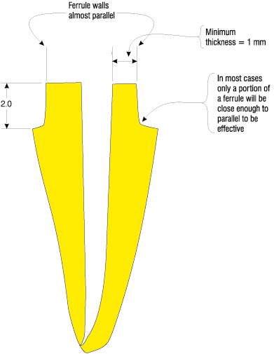
The importance of ferrule, its design and execution cannot be ignored. A recent in vitro study by Freeman and others31 compared leakage under post-retained composite cores and cast gold post cores and the number of cycles until a strain gage detected movement between the artificial crown and the root. Movement between crown and root was measured very early in the experiment, on average after only a few hundred cycles. In the experimental design, tooth preparation "extended 1 mm apical to the core-tooth interface, thereby providing a 1 mm ferrule on tooth structure."31 In reality, when the teeth were sectioned and the margins examined, there was little or no ferrule present. The cross-sections effectively showed a chamfered or bevelled margin that was far from parallel. Both Barkhordar32 and Sorensen33 have shown the importance of at least 1 mm of almost parallel wall preparation. The study by Freeman and others highlights the misconceptions as to what constitutes a ferrule. Given the realities of intraoral tooth preparation, a minimum of 2 mm of preparation length on solid tooth is recommended to ensure an adequate length of parallel wall to achieve an effective ferrule (Fig. 2).
Fig. 2 Ferrule criteria and dimensions
Posts have one purpose, to retain a build-up on a tooth. There is compelling evidence that they do not strengthen teeth.34-36 The use of certain post designs can predispose them to catastrophic failure, as shown by Sorensen and Engelman.33 Based on this premise, posts function primarily in tension. A relatively small diameter post has ample strength in tension to retain any crown. The rather loose adaptation of a small diameter post in a canal does not reduce retention, as shown by Chan and others.37 Standlee and others38 showed that post diameter was not a major factor in retention and the parallel-sided para post was more retentive than a smooth-tapered dowel design. In situations where an artificial crown breaks off and either shears off the post or dislodges it, there is at least the potential for retreatment. Utilization of larger posts requires the removal of additional tooth structure and weakens the tooth,39-41 increasing the risk of catastrophic failure and of having a tooth that is unrestorable.
Passive metal posts include parallel, tapered, and custom posts. Weine42 showed a 94% success rate with smooth-tapered posts over 10 years. In a study of 1,273 teeth restored a minimum of one year, Sorensen and Martinoff43 showed a 97% success rate for any post crown restoration in which the post length equalled or exceeded the crown length. Torbjorner and others44 reviewed almost 800 posts after four years, and parallel posts showed half the failure rate of custom-cast tapered posts. Post length is key and ideally should extend into the root past the crown margin by an amount equal to the length of the crown. Conservation of dentin is critical in post placement and dictates a post diameter that requires minimal canal instrumentation. Parallel post designs offer increased retention over tapered designs.37,38
Active post designs rely on some form of mechanical engagement of cutting flutes into dentin to gain increased retention. One active post design, the Flexi-Post by EDS, purports to provide the retention of an active post while avoiding the potential stress on the root of a conventional solid active post design. As pointed out by Manning and others,45 for the Flexi-Post design, the change in diameter provided by the slot in the post is minimal. The post compresses to an ellipse whose greatest width is almost equal to the original diameter. Standlee and Caputo46 showed Flexi-Posts generated significant stress levels comparable with other active post designs. While the additional retention of an active post has appeal, any active post design induces more stress into a root than a passive design.
There has been a lot of interest in carbon fibre post systems, specifically the composipost. These claims include ease of removal for retreatment (apparently valid), a modulus of elasticity (Young's) close to natural tooth, which decreases the risk of stress concentrations, and a high level of retention because the post bonds to the resin system used to retain it in the root. Sidoli and others25 showed in vivo crowns retained with composiposts failed under 63% of the load required to break para-post retained crowns. Purton and Love47 showed bonded para-posts to have 168% of the retention of carbon fibre posts. The suggestion that a Young's modulus close to tooth is an advantage is questionable when the coronal restoration will be a rigid cast metal or ceramic crown. The increased flexibility in the coronal and radicular tooth structure resulting from these posts should cause increased, not decreased, stress concentrations at the crown margins. Despite the preceding, Fredriksson48 and others showed an almost 100% success rate in a retrospective study of 236 teeth restored with carbon fibre posts for over two years. Carbon fibre posts may offer promise, but with conflicting evidence and limited clinical trials, they cannot be recommended for routine use. The clinical evidence is clear in both in vivo and in vitro studies that adequately designed passive posts deliver highly predictable results. Passive, small diameter, parallel post designs are predictable and simple to use and are the design of choice.
With a passive round post design as a starting point, the question is whether to use a cast post core or a preformed post-retained amalgam or composite build-up.
A cast post design is indicated where alignment of the proposed crown is significantly different from the inclination of the canal, which is often the case with anterior teeth. With most anterior, and some bicuspid teeth, there is also inadequate room for sufficient bulk of build-up material around the post to provide a solid unit. Thus for most anterior teeth and small bicuspid teeth requiring a post, the choice is a cast post core design. When used, a cast post core should utilize a high-strength type IV gold alloy or a similar high-strength non-precious alloy. Preformed posts with a direct build-up work very well in posterior teeth where there is room for sufficient bulk of build-up material. Canal angulation is infrequently a problem. Preformed posts with an amalgam build-up are often more conservative of tooth structure than cast gold in posterior teeth. They are generally less expensive and quicker to fabricate.
With preformed posts, stainless steel (SS) posts are stronger for any given size than titanium posts and they are more radiopaque. This radiopacity is an advantage, making a post easier to identify clinically. The Parapost system by Whaledent is currently the only passive, parallel, SS preformed post series on the market available in small diameters. The system provides 0.9, 1, 1.14 and 1.25 mm diameter posts in addition to larger diameters.
Where a preformed post is used to retain a composite or amalgam build-up, the ability of the build-up material to attach itself to the post is important. Chang and Millstein49 tested the retention of Paraposts and Unity posts by Whaledent, and the Flexi-post by EDS with two types of composite build-up as well as amalgam. They found that the amalgam core was significantly more retentive than either composite core. Their results show the importance of the retentive features built into a post head. When trimming preformed posts, it is essential that this feature be retained. Use of preformed posts without such features should be avoided.
From the perspective of the ability to retrieve the post in the case of fracture or for orthograde endodontic retreatment, the use of a resin cement is contraindicated. In addition, the ability of resin cements to provide long-term resistance to coronal leakage is dependent not only on the longevity of the dentin bond but also on the bond to the post. Both are unproven. There is little or no evidence that the increased retention offered by these cements is a factor in clinical success where adequate post length can be obtained. In fact, Standlee and Caputo50 warn that too much retention may predispose a tooth to fracture. ZnPO4, and resin modified glass ionomer cements such as vitremer luting, offer adequate retention and resistance to leakage and simplify post removal. Pure glass ionomer cements should work as well but are sensitive to moisture or the lack of it in a canal when setting. The use of resin cements should be reserved for cases outside of these criteria where adequate post length and retention are not available.
It is possible to disturb the apical seal during post-hole preparation. A 4 to 5 mm apical seal of gutta-percha is recommended based on research by several authors.51-53 Mattison,51 Camp,52 Suchina and Ludington,54 and Haddix and others55 reviewed a range of techniques for post hole preparation and the removal of gutta-percha. Mattison and others, Camp and Todd, and Suchina and Ludington found little difference between mechanical removal with Gates-Glidden burs and removal with a hot plugger, while Haddix and others found that removal with a warm plugger produced the least leakage. Gates-Glidden burs offer a simple and predictable method for the removal of gutta-percha.
The restoration of anterior teeth needs better criteria on which to base restorative decisions. Even knowing when to place a crown on an anterior tooth is very hard to determine. The issue of leakage with endodontically treated teeth is of concern: how much leakage is too much? Does the degradation of dentin bonding agents over time present a concern for clinically significant leakage under build-ups or along posts? This paper does not provide guidelines for the restoration of compromised endodontically treated teeth nor indicate how to restore structural integrity to existing crowns that have had an endodontic access placed through them. Both areas need research and guidelines to aid the clinician in restoring these teeth.
When restoring an endodontically treated tooth, the first step is to assess the level of predictability involved in the restoration. If the tooth meets the previously outlined criteria, then the following approach is recommended.
On anterior teeth with intact crowns, simply closing the access with composite is as successful as placing a crown. Where significant coronal destruction has occurred, use a crown with a cast post core.
A crown is indicated on all endodontically treated posterior teeth. In preparing the tooth, parallel ferrule walls are essential and should be a minimum of 2 mm long apico-coronally. In addition, the thickness of the remaining dentin should be no less than 1 mm on the buccal and lingual wall areas, and optimally interproximally as well.
For the build-up use amalgam, and where inadequate pulp chamber depth remains to retain the build-up, a preformed post should be placed. On some posterior teeth such as small upper first bicuspids, cast gold post cores will be preferable to amalgam where tooth size prevents adequate bulk of build-up around the post.
Where a post is needed, use a small diameter, passive round post requiring a minimum of dentin removal and use a post length that extends into the root past the crown margin by the length of the crown. Cement the post with ZnPO4 or a hybrid resin/glass ionomer cement, leaving an apical seal of 4 mm of gutta-percha.
Dr. Alex McLean is in private general practice in Kamloops, British Columbia.
Reprint requests to: Dr. Alex McLean, 201-418 St. Paul St., Kamloops, BC V2C 2J6.
The author has no declared financial interest in any company manufacturing the types of products mentioned in this article.
References
1. McLean AGR. Criteria for the predictably restorable endodontically treated tooth. J Can Dent Assoc 1998; 64:652-6.
2. Bullard RH, Leinfelder KF, Russell CM. Effect of coefficient of thermal expansion on microleakage. JADA 1998; 116:871-4.
3. Proprietary data provided by Williams, Amherst, NY.
4. Xu HC, Liu WY, Wang T. Measurement of thermal expansion coefficient of human teeth. Aust Dent J 1989; 34:530-5.
5. Mahler DB, Bryant RW. Microleakage of amalgam alloys: An update. JADA 1996; 127:1351-6.
6. Christensen GJ. Should you and can you afford to bond amalgams? JADA 1994; 125:1381-2.
7. Santos AC, Meiers JC. Fracture resistance of premolars with MOD amalgam restorations lined with amalgam bond. Oper Dent 1994;19:2-6.
8. Bonilla E, White SN. Fatigue of resin-bonded amalgam restorations. Oper Dent 1996; 21:122-6.
9. Meiers JC, Turner EW. Microleakage of dentin/amalgam alloy bonding agents: Results after 1 year. Oper Dent 1998; 23:30-3.
10. Burrow MF, Satoh M, Tagami J. Dentin bond durability after three years using a dentin bonding agent with and without printing. Dent Mater 1996; 12:302-7.
11. Proprietary data provided by Essential Dental Systems.
12. Proprietary data provided by Bisco Dental.
13. Sakaguchi RL, Sasik CT, Bunczak MA, Douglas WH. Strain gauge method for measuring polymerization contraction of composite restoratives. J Dent 1991; 19:312-6.
14. Arikawa H, Kuwahata H, Seki H, Kanie T, Fujii K, Inoue K. Deterioration of mechanical properties of composite resins. Dent Mater J 1995; 14:78-83.
15. Kovarik RE, Breeding LC, Caughman WF. Fatigue life of three core materials under simulated chewing conditions. J Prosthet Dent 1992; 68:584-90.
16. Carlson TJ, Naguib EA, Cochran MA, Lund MR. A comparison of glass ionomer cements used to repair cast restorations. Oper Dent 1990; 15:162-6.
17. Craig RG. (1997) Restorative dental materials. CV Mosby St. Louis.
18. Bryant RW, Mahler DB. Modulus of elasticity in bending of composites and amalgams. J Prosthet Dent 1986; 56:243-8.
19. Cohen BI, Pagnillo M, Musikant BL, Deutsch AS, Cofrancesco G. Comparison of the Young's modulus for six reinforced dental materials. Oral Health 1997; 87:47-8.
20. Huang TJ, Schilder H, Nathanson D. Effects of moisture content and endodontic treatment on some mechanical properties of human dentin. J Endod 1992; 18:209-15.
21. Caputo AA, Standlee JP. Pins and posts - why, when and how. Dent Clin North Am 1976; 20:299-311.
22. Nayyar A, Walton RE, Leonard LA. An amalgam coronal-radicular dowel and core technique for endodontically treated posterior teeth. J Prosthet Dent 1980; 43:511-5.
23. Kane JJ, Burgess JO, Summitt JB. Fracture resistance of amalgam coronal-radicular restorations. J Prosthet Dent 1990; 63:607-13.
24. Sorensen JA, Martinoff JT. Intracoronal reinforcement and coronal coverage: A study of endodontically treated teeth. J Prosthet Dent 1984; 51:780-4.
25. Sidoli GE, King PA, Setchell DJ. An in vitro evaluation of a carbon fibre-based post and core system. J Prosthet Dent 1997; 78:5-9.
26. Torabinejad M, Ung B, Kettering JD. In vitro bacterial penetration of coronally unsealed endodontically treated teeth. J Endod 1990; 16:566-9.
27. Swanson K, Madison S. An evaluation of coronal microleakage in endodontically treated teeth. Part I: Time periods. J Endod 1987; 13:56-9.
28. Madison S, Swanson K. An evaluation of coronal microleakage in endodontically treated teeth. Part II: Sealer types. J Endod 1987; 13:109-12.
29. Khayat A, Lee SJ, Torabinejad M. Human saliva penetration of coronally unsealed obturated root canals. J Endod 1993; 19:458-61.
30. Ray HA, Trope M. Periapical status of endodontically treated teeth in relation to the technical quality of the root filling and the coronal restoration. Int Endod J 1995; 28:12-8.
31. Freeman MA, Nicholls JI, Kydd WL, Harrington GW. Leakage associated with load fatigue-induced preliminary failure of full crowns placed over three different post and core systems. J Endod 1998; 24:26-32.
32. Barkhordar RA, Radke R, Abbasi J. Effect of metal collars on resistance of endodontically treated teeth to root fracture. J Prosthet Dent 1989; 61:676-8.
33. Sorensen JA, Engelman MJ. Ferrule design and fracture resistance of endodontically treated teeth. J Prosthet Dent 1990; 63:529-36.
34. Guzy GE, Nicholls JI. In vitro comparison of intact endodontically treated teeth with and without endo-post reinforcement. J Prosthet Dent 1979; 42:39-44.
35. Assif D, Bitenski A, Pilo R, Oren E. Effect of post design on resistance to fracture of endodontically treated teeth with complete crowns. J Prosthet Dent 1993; 69:36-40.
36. Trope M, Maltz DO, Tronstad L. Resistance to fracture of restored endodontically treated teeth. Endod Dent Traumatol 1985; 1:108-11.
37. Chan FW, Harcourt JK, Brockhurst PJ. The effect of post adaptation in the root canal on retention of posts cemented with various cements.
38. Standlee JP, Caputo AA, Hanson EC. Retention of endodontic dowels: Effect of cement, dowel length, diameter, and design. J Prosthet Dent 1978; 39:400-5.
39. Leary JM, Aquilino SA, Svare CW. An evaluation of post length within the elastic limits of dentin. J Prosthet Dent 1987; 57:277-81.
40. Hunter AJ, Feiglin B, Williams JF. Effect of post placement on endodontically treated teeth. J Prosthet Dent 1989; 62:166-72.
41. Tjan AH, Whang SB. Resistance to root fracture on dowel channels with various thicknesses of buccal dentin walls. J Prosthet Dent 1985; 53:496-500.
42. Weine FS, Wax AH, Wenckus CS. Retrospective study of tapered, smooth post systems in place for 10 years or more. J Endod 1991; 17:293-7.
43. Sorensen JA, Martinoff JT. Clinically significant factors in dowel design. J Prosthet Dent 1984; 52:28-35.
44. Torbjorner A, Karlsson S, Odman PA. Survival rate and failure characteristics for two post designs. J Prosthet Dent 1995; 73:439-44.
45. Manning KE, Yu DC, Yu HC, Kwan EW. Factors to consider for predictable post and core build-ups of endodontically treated teeth. Part II: Clinical application of basic concepts. J Can Dent Assoc 1995; 61:696-707.
46. Standlee JP, Caputo AA. The retentive and stress distributing properties of split threaded endodontic dowels. J Prosthet Dent 1992; 68:436-42.
47. Purton DG, Love RM. Rigidity and retention of carbon fibre versus stainless steel root canal posts. Int Endod J 1996; 29:262-5.
48. Fredriksson M, Astback J, Pamenius M, Arvidson K. A retrospective study of 236 patients with teeth restored by carbon fibre reinforced epoxy resin posts. J Prosthet Dent 1998; 80:151-7.
49. Chang WC, Millstein PL. Effect of design of prefabricated post heads on core materials. J Prosthet Dent 1993; 69:475-82.
50. Standlee JP, Caputo AA. Endodontic dowel retention with resinous cements. J Prosthet Dent 1992; 68:913-7.
51. Mattison GD, Delivanis PD, Thacker RW, Hassel KJ. Effect of post preparation on the apical seal. J Prosthet Dent 1984; 51:785-9.
52. Camp LR, Todd MJ. The effect of dowel preparation on the apical seal of three common obturation techniques. J Prosthet Dent 1983; 50:664-6.
53. Goodacre CJ, Spolnik KJ. The prosthodontic management of endodontically treated teeth: A literature review. Part II: Maintaining the apical seal. J Prosthodont 1995; 4:51-3.
54. Suchina JA, Ludington JR. Dowel space preparation and the apical seal. J Endod 1985; 11:11-7.
55. Haddix JE, Mattison GD, Shulman CA, Pink FE. Post preparation techniques and their effect on the apical seal. J Prosthet Dent 1990; 65:515-9.