![]()
![]()
Résultats de l’exposition chirurgicale, de la liaison et de l’éruption de 82 canines maxillaires incluses
Marco F. Caminiti, DDS, FRCD(C)
George K.B. Sandor, DDS, MD, FRCD(C), FRSC(C), FACS
Claudia Giambattistini, DDS
Bryan Tompson, DDS
SOMMAIRE
Historique
Grâce au perfectionnement des techniques de liaison et des matériaux, on peut poser des
attachements solides aux dents en position ectopique. Dans cette étude prospective, on
évalue les résultats de l’éruption orthodontique forcée de canines incluses se
trouvant dans les positions palatine et labiale.
Méthodes
L’étude a porté sur 82 canines maxillaires incluses de 54 patients et a duré de 18
ŕ 30 mois aprčs leur exposition. Aprčs les avoir exposées au moyen d’un lambeau
palatin ou d’un lambeau buccal remis en position apicale, on a, ŕ l’aide
d’un ciment en résine orthodontique polymérisée, lié ŕ chaque dent incluse un
crochet de traction orthodontique auquel était attachée une chaînette ŕ ligature.
Puis, sur le champ opératoire, on a placé un panse-ment parodontal pour quelque temps.
Résultats
Toutes les dents ont fait éruption. Parmi les complications éprouvées, mentionnons :
l’échec de la premičre liaison au moment de la chirurgie, ce qui a exigé une
nouvelle liaison; une perte prématurée de la liaison au moment d’enlever le
pansement; une perte de la liaison des attachements pendant l’éruption
orthodontique. Il n’y a eu ni infections, ni éruptions manquées, ni ankyloses, ni
résorptions, ni pertes parodontales (des poches plus profondes que 3 mm) associées aux
dents exposées. On a remarqué une gencive attachée de moins de 3 mm pour seulement deux
des canines en position buccale (9 p. 100).
Conclusion
L’éruption orthodontique forcée des canines maxillaires incluses ŕ l’aide
d’un crochet de traction bien lié et d’une chaînette ŕ ligature,
conjointement avec un lambeau palatin ou un lambeau labial remis en position apical, donne
des résultats prévisibles avec peu de complications.
Mots clés MeSH: cuspid/surgery; orthodontics, corrective; tooth, impacted/therapy, outcomes.
[ Introduction | Materials and Methods | Results | Discussion | Clinical Significance | Conclusion | Acknowledgements | References ]
The purpose of this study is to evaluate the success of a one-step technique for exposing and guiding into occlusion, impacted maxillary canines. The technique involves using the most recently available bonding materials and attachment devices, in conjunction with a consistent surgical approach. Teeth may be impacted or erupt ectopically for a variety of reasons. Hereditary factors, lack of space, persistence of primary canines, a true ectopic path of eruption, reduced root length and aplasia of lateral incisors are a few of the factors cited.1
Ericson and Kurol 2 reported that, in terms of the frequency of impaction, the maxillary permanent canine ranks second only to the third molar, with a prevalence of approximately 2% in the general population. Impacted canines are positioned palatally 85% of the time. The frequency of impaction is three times greater in females than males.3 Some patients with craniofacial anomalies, such as cleidocranial dysplasia and cretinism, have multiple unerupted teeth. These groups present with specific problems in relation to the timing and nature of treatment.4,5
The location of the impacted tooth determines the type of surgical approach. In general, there are three steps to clinical localization.6 Visual inspection and digital palpation are the first two steps, while radiographic examination is the third and most critical step. Periapical, occlusal, cephalometric, posterior-anterior and panoramic radiographs, as well as polytomography have all been used to localize impacted canines.3,7,8
Periapical radiographs give a good indication of the labial- palatal location of the canine, relative to the incisors, by means of the parallax technique (or the Tube- shift technique/Clark rule). The vertex occlusal is the most accurate of the occlusal films for determining the position of the canine, relative to the midline.9 Superimposition of structures may make interpretation of this radiograph difficult and unreliable. The lateral cephalogram reveals the anterior-posterior position, inclination and vertical location of the canine. However a 45° oblique cephalometric film may be more accurate, due to reduced double image formation.9
The panoramic radiograph, in conjunction with the periapical, or the maxillary oblique radiograph, is the preferred method to localize impacted canines.2,6 The image of a tooth located palatal to the dental arch is relatively magnified, while that of a buccally placed tooth appears diminished.10 In a study of 109 ectopic canines, researchers could predict the palatal location of impacted canines with an 80% specificity using panoramic radiographs.10
Various treatment modalities have been proposed to avoid the complications associated with impacted canines.3,11,12 These complications include: internal or external resorption; infection associated with partial eruption; loss of arch length; and most commonly, the resorption of the roots of lateral incisors.2,
Canine impactions may be prevented through early diagnosis. According to Ericson and Kurol,3 if a maxillary permanent canine appears to be erupting ectopically or not erupting at all, the extraction of the primary canine is recommended in the 10-13 age group. These investigators demonstrated the importance of early diagnosis. In 78% of palatally erupting canines, a normal path of eruption was established within 12 months of removal of the primary canine.
Theofanatos and coworkers 13 reported that 91% of ectopically erupting canines came into proper occlusion if the canine crown was distal to the midline of the lateral incisor at the time of removal of the deciduous canine. If, however, the crown was mesial to the midline of the lateral incisor root, spontaneous eruption occurred in only 64% of cases.
If prevention is not successful, intervention is warranted. According to Proffitt,14 there are three categories of problems when dealing with an impacted tooth: surgical exposure; attachment to the tooth and; orthodontic mechanics to bring the tooth into the arch. Bishara 15 advocates the surgical exposure of the impacted canine with no orthodontic traction, only when the tooth has a correct axial inclination. Surgically exposed teeth rarely erupt into a created space, without aid, especially once root formation is complete.3
Numerous surgical techniques have been described to expose impacted teeth.6,9,11,16-19 Earlier techniques were limited by the materials available to bond to the tooth. Wire ligatures and window flaps were originally proposed.15,17 These techniques are associated with poor periodontal healing, with loss of attachment, gingival recession, and gingival inflammation. 15,20 Newer flap designs have resulted in better postoperative periodontal .19,21-23
As bonding techniques developed in orthodontics, surgical procedures were modified, since less of the impacted crown now needed to be exposed. Newer ortho-dontic resins have increased bond strengths, decreased setting times and are light cured. Light cured bonding agents compare favorably with those that are chemically cured.24
The application of etch and bonding agents has been improved by making these less sensitive to the moisture of the surgical site. Smaller brackets and improved wires have led to more predictable results. Newer surgical dressings are light cured, have a palliative effect, maintain their position well, are easier to place, adapt flaps better and decrease hematoma formation.7,25
This study included 54 ortho-dontic patients (31 female, 23 male) who were followed for 18-30 months after the surgical exposure of canines. The patients were 12 to 16 years old (mean female=12.7, male=14). A total of 82 impacted maxillary canines were treated according to a standard protocol. Sixty teeth were palatally impacted and 22 were labially impacted. At the time of surgical exposure, all the teeth had at least one-third to two-thirds of root formation completed. All the crowns of the impacted teeth were covered by bone.
Prior to surgery, the position of the impacted tooth was determined clinically (by palpation) and radiographically (panoramic, lateral cephalogram, two periapicals and maxillary occlusal films).9,16,26
For bilateral palatally impacted canines, a full thickness mucope-riosteal flap was raised from the mesiopalatal aspect of the second premolars. In unilateral cases, the flap extended to the mesiopalatal aspect of the contralateral lateral incisor (Fig. 1).
For labially impacted canines, a full thickness mucoperiosteal flap was raised, initially without vertical incisions, from the mesiobuccal aspect of the second premolar to the distolabial aspect of the lateral incisor. Vertical releasing incisions were placed mesially and distally. Sufficient bone was removed from around the crown in order to place a bonded orthodontic appliance. An apically positioned flap was fashioned if the tooth was coronally placed (Fig. 2). For deeper impactions, a closed eruption technique was employed.
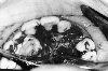
Fig 1:Extent of a unilateral
palatal flap for
the exposure of a high impacted canine (*).
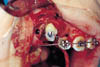
Fig 2a:Labially positioned
impacted cuspid exposed
with orthodontic attachment in place.
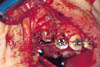
Fig 2b:Apically repositioned flap
sutured into place at
the site of a labially impacted canine tooth.
The enamel was etched for 60 seconds using 37% phosphoric acid. At this point the field was kept as dry as possible using suction and gauze or bone wax. A light cured orthodontic resin cement (Unitek, Transbond XT) was used for bonding (light cured at 470 nm for 40 seconds). The appliance used was a bonded orthodontic traction hook with a ligation chain (Fig. 2) (TP Orthodontics).
If the primary canine was present, it was extracted and the wire was passed through the extraction socket (as described by Crescini 12 ). The ligation chain was wrapped around the existing labial archwire, or held in place within the periodontal dressing until the ortho-dontic appliance was placed.
The flap was then repositioned. Labial flaps were apically repositioned ensuring that the full amount of keratinized gingiva was maintained cervical to the crown. Palatal flaps were repositioned to their original site and windows were placed in the mucoperiosteum, for the more medially placed palatal canines, to allow for passage of the wire. Flaps were held in place by means of resorbable sutures.
Subsequently, a light cured periodontal dressing (Barricaide) was placed over the area (Fig. 3). The dressing assured patency of the window, aided flap immobilization, and provided protection and comfort for the patient.
Patients were then evaluated 7-14 days after surgery, when the dressing and sutures were removed. Clinical evaluation included assessment of bracket attachment, eruption status, gingival tissue response, recession, periodontal pocket depth and infection. A radiographic examination with one standard periapical film was performed to assess the status of adjacent structures, as well as the presence of root resorption, ankylosis or periodontal defects.
The patients then had orthodontic traction forces activated within seven to 21 days. At subsequent orthodontic appointments, the same clinical examination was performed with radiological evaluation occurring every three months. Progress was noted and complications were recorded.
All teeth were successfully guided into occlusion. Table I lists the number and type of complications that occurred from the time of the surgery through to the final alignment of the canines. The periods when complications occurred were classified as: surgical, post-operative and orthodontic.
Complications in the surgical stage consisted of failure of the initial bond at the time of surgery. One labial (4.5%) and two palatal (3.3%) canines were immediately rebonded. Bond failure was most often due to the difficulty of obtaining a dry field. Post-operatively, one palatal canine (1.6%) required rebonding when the packing was removed. During orthodontic traction, only one palatal and one labial canine required rebonding (neither had debonded previously). Clinically, there were no cases of infection, eruption failure, ankylosis, gingival inflammation, or gingival recession. Three labially placed teeth had pockets, greater than 3 mm, five months postoperatively (13.6%). Radiographically there was no evidence of resorption, bone defects or infection.
Once complete eruption was achieved, there were no periodontal defects or pockets greater than 3 mm. All sites had at least a 3 mm band of attached gingiva except for two labially placed canines which had a 1 mm band (9%). Orthodontic treatment continued uneventfully until debonding (Fig. 4).
| Table I | |||||||||||||||||||||||||||||||||||||||||||||
| The Nature and Timing Of Complications Presented By Eight Cases Whose Maxillary Canines Were Exposed. | |||||||||||||||||||||||||||||||||||||||||||||
|
|||||||||||||||||||||||||||||||||||||||||||||
|
During orthodontic treatment, it is particularly important to maintain a healthy band of keratinized gingiva around a labially positioned canine, otherwise the mobile tissue around the tooth may strip away from the crown and root surface leaving a periodontal defect.11,14,17,18,22 An adequate band of attached gingiva can be achieved by either apically repositioning a flap with attached gingiva, or by grafting keratinized gingiva from the palate to the site of exposure.
Boyd 20 demonstrated that in labially positioned canines, a 2–3 mm band of attached gingiva created by an apically repositioned flap is preferable to a window exposure with no attention to keratinized tissue. This technique results in a significant reduction of gingival recession, inflammation and loss of attachment.
Removing smaller amounts of overlying bone during surgery, results in reduced bone loss after orthodontic treatment.14,20,21 The importance of this finding has been disputed,11 especially in the case of palatal impactions, where bone removal may be less critical.
With appropriate handling of the soft tissues during the surgical phase and proper oral hygiene practices postoperatively, the exposed teeth were periodontally healthy in this study (Fig. 4). With respect to the management of the soft tissues, the results of the present study are supported by those of other investigators.11,17,18,20,22 The closed eruption technique that was used on the palatally impacted teeth and on some that were labially impacted, produced favorable results. Vermette and coworkers 19 comparing the closed eruption technique to that using apically repositioned flaps, found that closed eruption produced more favorable esthetics, less crown length exposure and a similar periodontal outcome.
Many methods of attaching “hardware” to teeth have been described.6,11,20,29 Originally, wire ligatures were placed around the crown of the impacted tooth but, this had the potential to upset the periodontal attachment.26 Boyd 23 compared wire ligation to bonding brackets on palatally impacted canines. In general, the wire ligated teeth had a greater incidence of non-eruption, ankylosis, external root resorption and loss of attached mucosa, due to the larger flaps required.
Before the development of bonding, retentive pins were drilled into the teeth. From these pins, wires were suspended to drag the teeth into occlusion. The risk of pulpal injury, limits the use of this technique today.14 A preferable approach is to expose an area of the tooth and bond a hook or button attachment at the time of surgery. With the advent of rapid light-curing orthodontic resins, which are as effective as self-cured resins,24 the surgeon is now in a position to place the attachment in the most ideal location.
Lewis 6 originally advocated a two-step approach for the attachment to the canine. First, the tooth was exposed and packed. Second, after three to eight weeks, the pack was removed by the orthodontist and the bracket was placed. Premature loss of the pack and closure of the defect often occurred prior to the orthodontic visit.
With the bracket attached to the tooth during the surgical procedure, the flap is repositioned over or around the exposed canine. The wire already attached to the bracket is then brought up to the arch-wire for stabilization and attachment. The use of a traction wire tunnelled through the extraction socket was found to be effective, in this study and in others.12,19
Ideally, mechanical traction should be activated immediately after surgery and the force should be applied to an existing fixed 14 or removable appliance.11 Traction in this study was activated within 7-14 days post operatively. This delay did not produce any long term orthodontic or periodontal complications. Several wires or elastomeric threads were used during this traction phase.
Other techniques to guide eruption include: special alignment springs called Ballista springs (if brackets are already in place in the buccal segments); coil springs; power chains; or loops of ligature wire extending from the canine to the archwire.1,9,15,20 Magnets have recently been used to produce eruptive forces.28,29
Complications in treating impacted canines include failure to erupt,15 periodontal defects,21,22,30 bond failure,15,20,23 and ankylosis. 3,20,23 The effect of ankylosis is to prevent tooth eruption. This may cause the anchoring teeth, on the archwire, to tip into the space created for the canine. Ankylosis has also been implicated in some cases where the canine initially moved and then suddenly ceased to erupt. Luxation of the ankylosed tooth has been a recommended treatment in this case however, success is unpredictable.9
In this study, all of the 82 impacted canines treated, erupted. This success rate may be partly due to early diagnosis and to the age of the patients. It is recommended to treat this condition before the age of 20, to maximize the potential for success.31
The frequency of bond failure, the most common complication observed in this study, was minimal. Other studies have not addressed the problems of bracket detachment, which may not be a significant factor if the bracket is bonded well at the initial surgical procedure.
Where a dry field can’t be attained, the bonding of brackets is unpredictable. In this situation, alternative materials such as light cured and chemically cured glass ionomer cements (Fuji Ortho LC) or a 4-meta resin cement (C&B Metabond, Parkell), could be used. This study did not evaluate the relative effectiveness of such materials, although this could be the subject of a future study.
A rigid light cured periodontal dressing, such as the Barricaide used in this study, can be kept in place for up to two weeks postoperatively (Fig. 3). The dressing may be useful in maintaining the patency of the exposure of palatally impacted canines and in adapting an apically positioned labial flap. In this study, the one premature debonding observed at the time of follow-up was easily rebonded, because the dressing maintained a clean tunnel to the tooth.
Barricaide, a visible light cured periodontal dressing material, exhibits superior physical properties and ease of handling, as compared to standard, chemically cured, dressing materials. It maintains its tensile strength even when immersed in water. It can be placed in the embrasure spaces to increase its retention, although etching adjacent enamel and applying a bonding agent has also been proposed.25
In an animal histologic study comparing different dressing materials, Barricaide, which was present for two weeks showed no deleterious effects on healing.32 In this study, the membrane was retained in all patients for the first postoperative week. The good soft tissue healing, lack of infection and hematoma, as well as flap adaptation made the dressing a helpful adjunct.
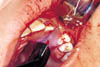
Fig 3a:Adaptation of light cured
periodontal dressing to
close a flap of a palatally impacted canine.
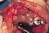
Fig 3b:Light cured periodontal
dressing stabilizing
an apically repositioned flap.
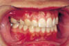
Fig 4:Successfully erupted tooth 13
with health periodontal tissues.
The tooth had been labially positioned.
• The exposure of impacted canines with the use of a palatal flap or an apically
repositioned labial flap allows predictable orthodontic eruption with minimal
complications.
• Newer light-cured bonding agents provide quick and dependable bonded attachments to
enamel.
• A prefabricated orthodontic attachment allows ease of placement.
• Light-cured rigid elastic packing materials produce favorable soft tissue results.
Dr. Caminiti is the former chief resident, department of oral and maxillofacial
surgery, University of Toronto and The Toronto Hospital. He is a clinical fellow at The
Centre for Research in Education, University of Toronto, Faculty of Medicine.
Dr. Sandor is assistant professor, oral and maxillofacial surgery, University of Toronto; coordinator, oral and maxillofacial surgery, The Hospital for Sick Children and Bloorview MacMillan Centre; staff, oral and maxillofacial surgery, The Toronto Hospital, Toronto, Ontario.
Dr. Giambattistini is formerly resident, department of orthodontics, University of Toronto, Toronto, Ontario.
Dr. Tompson is head, division of orthodontics, The Hospital for Sick Children, Toronto, Ontario.
Reprint requests to: Dr. George K.B. Sandor, Department of Dentistry, Hospital for Sick
Children, 555 University Ave., Toronto ON M5G 1X8
| 1. | Jacoby H. The etiology of maxillary canine impactions. Am J Orthod 1983; 84:125-32. | |
| 2. | Ericson S, Kurol J. Radiographic examination of ectopically erupting maxillary canines. Am J Orthod 1987; 91:483-92. | |
| 3. | Ericson S, Kurol J. Early treatment of palatally erupting maxillary canines by extraction of the primary canines. Eur J Orthod 1988; 10:283-95. | |
| 4. | Davies TM, Lewis DH, Gillbe GV. The surgical and orthodontic management of unerupted teeth in Cleidocranial dysostosis. Br J Orthod 1987; 14:43-7. | |
| 5. | Trimble LD, West RA, McNeill RW. Cleidocranial dysplasia: comprehensive treatment of the dentofacial abnormalities. J Am Dent Assoc 1982; 105:661-6. | |
| 6. | Lewis PD. Preorthodontic surgery in the treatment of impacted canines. Am J Orthod 1971; 60:382-97 | |
| 7. | Holmes A, Nashed RR. Radiographic localization of canines in general dental practice. Dent Update 1990; 17:29-34. | |
| 8. | Kuftinec MM, Stom D, Shapira Y. The impacted maxillary canine I. A review of concepts. ASDC J Dent Child 1995; 62:317-24. | |
| 9. | Kasander T. The impacted canine: diagnosis and treatment, part I. J Gen Orthod 1994; 5:13-22,27. | |
| 10. | Fox NA, Fletcher GA, Horner K. Localising maxillary canines using dental panoramic tomography. Br Dent J 1995; 179:416-20. | |
| 11. | MacDonald F, Yap W. The surgical exposure and application of direct traction of unerupted teeth. Am J Orthod 1986; 89:331-40. | |
| 12. | Crescini A, Clauser C, Giogetti R, Pini G. Tunnel traction of infraosseous impacted maxillary canines: a three year periodontal follow up. Am J Orthod Dentofacial Orthop 1994; 105:61-72. | |
| 13. | Theofanatos GD, Zavras AI, Turner IM. Periodontal considerations in the treatment of maxillary impacted cuspids. J Clin Pediatr Dent 1994; 18:245-52. | |
| 14. | Proffitt WR. Contemporary Orthodontics, St. Louis: CV Mosby, 1986, pp 408-11. | |
| 15. | Bishara SE. Impacted maxillary canines: A review. Am J Orthod Dentofacial Orthop 1992; 101:159-71 | |
| 16. | Bishara SE, Kommer DD, McNeil MH et al. Management of impacted canines. Am J Orthod 1976; 69:371-87. | |
| 17. | McBride LJ. Traction, a surgical-orthodontic procedure. Am J Orthod 1979; 76:287-99. | |
| 18. | Wisth PJ, Nordeval K, Boe OE. Comparison of surgical methods in combined surgical-orthodontic correction of impacted maxillary canines. Acta Odontol Scand 1976; 34:53-57. | |
| 19. | Vermette ME, Kokich VG, Kennedy DB. Uncovering labially impacted teeth: apically positioned flap and closed-eruption techniques. Angle Orthod 1995; 65:23-34. | |
| 20. | Boyd R. Clinical assessment of injuries in orthodontic movement of impacted teeth II: Surgical recommendations. Am J Orthod 1984; 86:407-18. | |
| 21. | Heaney TG, Atherton JD. Periodontal problems associated with the surgical exposure of unerupted teeth. Br J Orthod 1976; 3:79-84. | |
| 22. | Vanarsdall RL, Corn H. Soft tissue management of labially positioned unerupted teeth. Am J Orthod 1977; 72:53-64. | |
| 23. | Boyd R. Clinical assessment of injuries in orthodontic movement of impacted teeth I: Methods of attachment. Am J Orthod 1982; 82:478-86. | |
| 24. | Wang WN, Meng CL. A study of bond strength between light and self-cured orthodontic resin. Am J Orthod Dentofacial Orthop 1992; 101:350-4. | |
| 25. | von Fraunhofer JA, Argyropoulos DC. Physical properties of a periodontal dressing material. Am J Dent 1992; 5:266-8. | |
| 26. | Kuftinec MM, Stom D, Shapira Y. The impacted maxillary canine II: Clinical approaches and solutions. ASDC J Dent Child 1995; 62:325-34. | |
| 27. | Ferguson JW. The use of visible light cured periodontal dressing after surgical exposure of palatal canines. Dent Update 1992; 19:380-2,4 | |
| 28. | Sandler PH, Fearne J. Unerupted Incisors: a case report illustrating an attractive solution. J Int Assoc Dent Child 1990; 20:22-25. | |
| 29. | Darendeliler MA, Friedli JM. Treatment of an impacted canine with magnets. J Clin Orthod 1994; 28:639-43. | |
| 30. | Kohavi D, Becker A, Zilberman Y. Surgical exposure, orthodontic movement, and final tooth position as factors in periodontal breakdown of treated palatally impacted canines. Am J Orthod 1984; 85:72-7. | |
| 31. | Nordenram A. Impacted maxillary canines - a study of surgically treated patients over twenty years of age. Swed Dent J 1987; 11:153-8. | |
| 32. | Smeekens JP, Maltha JC, Renggli HH. Histological evaluation of surgically treated oral tissues after application of a photocuring periodontal dressing material. An animal study. J Clin Periodontolol 1992; 19:641-5. |