S
upporting
Y
our
P
ractice
Management of “HiddenCaries”:
ACase of Severe
Pre-eruptive Intracoronal Resorption
The following is based on a research article originally published
in the “Clinical Reports” section of
jcda.ca
—CDA’s online, open
access scholarly publication that features articles indexed in
Medline, Journal CitationReports and ScienceCitation Index.
TimucinAri
DDS,PhD
Pre-eruptive intracoronal resorption (PEIR) appears as a radiolucent lesion in thecoronal
dentin, adjacent to thedentin-enamel junctionof unerupted teeth. AlthoughPEIR resembles
dental carieson radiographs, there is littlehistopathologicormicrobiologicevidence to
support that hypothesis.
Dr. TimucinAri, assistant professor in theorthondontics andpediatricdentistrydivisionat
SchulichDentistry, presents acaseof post-eruptivediagnosisof intracoronal resorption
withextensivedestructionof dentine. This case report emphasizes the importanceof early
detectionof the resorptiveprocess tominimize itspotentiallydestructivecapacity.
Casereport
A12-year-oldpatient reported toapediatricdental clinic. Hehadpreviouslyattended the
sameclinicat age8, but had failed toattendanyappointments since that time. His chief
complaintwas spontaneous, throbbingpainwithassociated swelling in the leftmandibular
area. Thechildhadbeenunable tochewon theaffected side for 12hours. Therewasno
historyof trauma, andmedical history revealedgoodgeneral health.
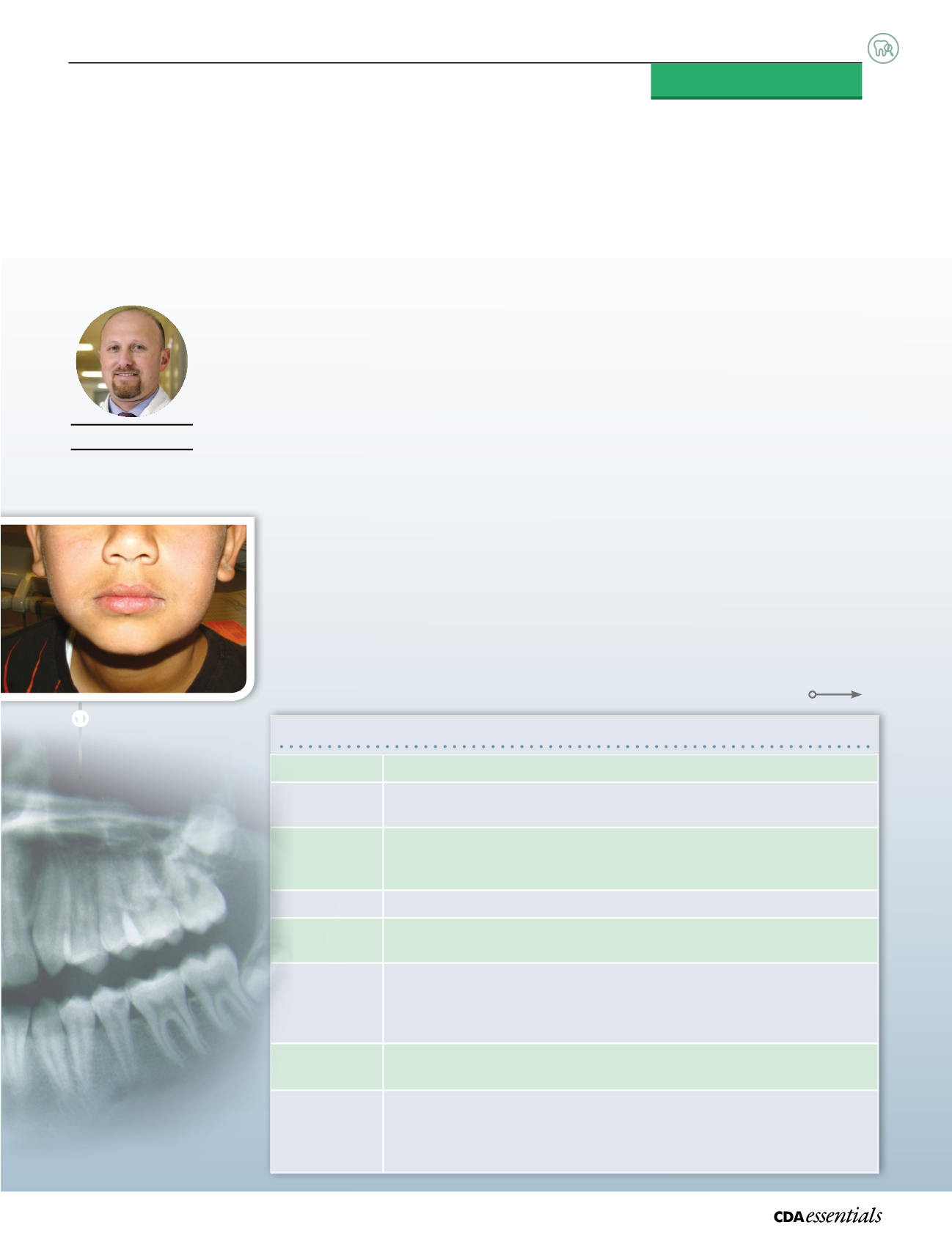
Duringextra-oral examination, adiffuse soft andmobile swellingwaspalpatedon the
left lateral surfaceof themandible, extending inferiorlydown to the lower border of the
mandible (
Fig.
➊
). Submandibular lymphglandsonboth sideswerepalpableand tender.
Clinical Summary
➊
➊
Swellingon the left lateral
surfaceof themandible.
Pre-eruptive intracoronal resorption: Presentation
Population
• noassociationwith sex, race,medical status, systemicdiseaseorfluoride supplementation
Prevalence
• 1.55% to6%, dependingon the typeandqualityof the radiographicexposureandageof patients
• Moreprevalentwhen thirdmolarsare included
Siteof
occurrence
• coronal dentin, adjacent to thedentin-enamel junctionof unerupted teeth
• usuallya single tooth isaffected
• almosthalf of the lesionsextend tomore than2/3of the thicknessof thedentin
Symptoms
• usuallyminimal or absent
Signs
• large lesionmaybepresent in the coronal dentinadjacent to thedentin-enamel junction
• radiographicappearanceof the radiolucency
Pathogenesis
• unclear
• ithasbeenhypothesized that local factors (e.g., damage to the reducedenamel epitheliumor
unerupted teeth)mayallow invasionof cells from surroundingbony tissue to the surfaceof the
developing tooth
Detection
• most lesions remainundetectedbecauseof the lackof symptomsand theproblems involved in
achievinganoptimumview inbite-wing radiographsof themixeddentition
Evolution
• progressionusually slowsbefore the tootherupts
• mostdefects remainadjacent to thedentin-enamel junction, rarelyextending into thepulp
• once toothhasemerged into theoral cavity, conditionsbecomeappropriate for rapid
developmentof caries
33
Volume2 Issue6
|


