|
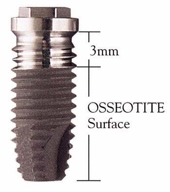
| Figure 1: Osseotite implant: 3 mm polished coronal surface for soft tissue health and patented Osseotite acid-etched surface for increased mechanical interlocking with bone. |
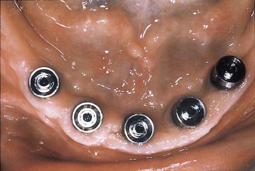 |
| Figure 2a: Five standard abutments in place to support a long-span fixed bridge with bilateral distal cantilevers (Group A). |
 |
| Figure 2b: Space designed for hygiene of a long-span fixed bridge with bilateral distal cantilevers (Group A). |
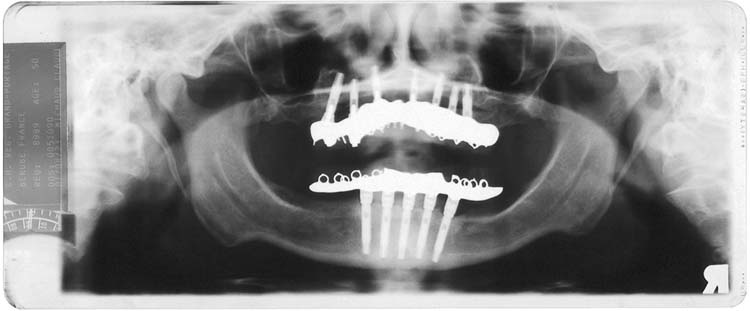 |
| Figure 2c: Panoramic radiograph of a mandibular long-span fixed bridge with bilateral distal cantilevers (Group A), with a maxillary fixed-removable implant-supported prosthesis. |
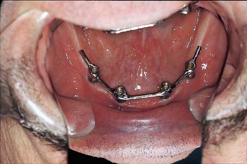 |
| Figure 3a: Metal bar with bilateral distal cantilevered extensions (mirror image) supporting an overdenture (Group A). |
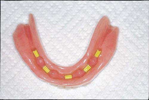 |
| Figure 3b: Lower complete denture with clip attachments for retention of an overdenture (Group A). |
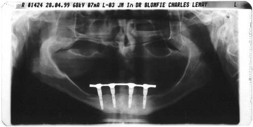 |
| Figure 3c: Panoramic radiograph with screw-retained metal on 4 implants supporting an overdenture (Group A). |
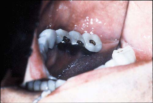 |
| Figure 4a: Three splinted screw-retained crowns (lingual view, mirror image of the lower left posterior region, Group C). |
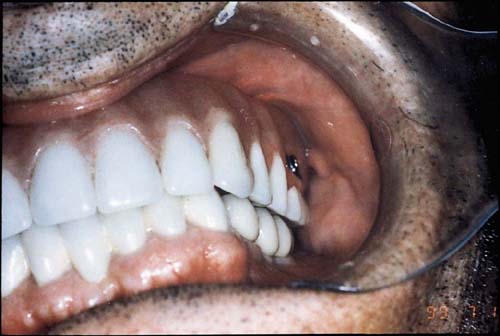 |
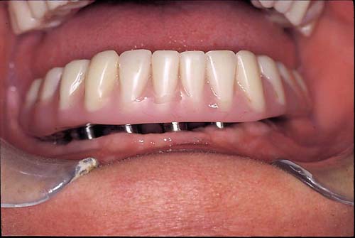
| Figure 4b: Buccal view of 3 splinted screw-retained lower left posterior crowns with a fixed-removable maxillary denture in place (Group C). |
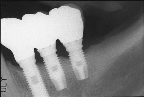 |
| Figure 4c: Periapical radiograph of 3 splinted screw-retained lower left posterior crowns (Group C). |