|
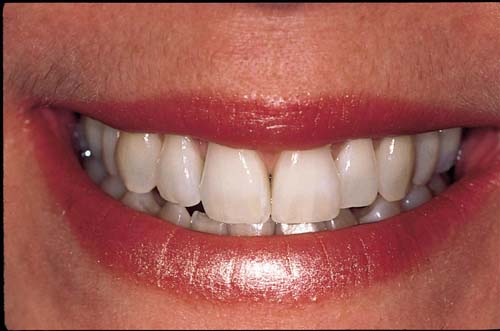
| Figure 1: The dental midline is oriented vertically in the face and is perpendicular to both the maxillary incisal curve and the lower lip curve, contributing to a pleasing visual symmetry. |
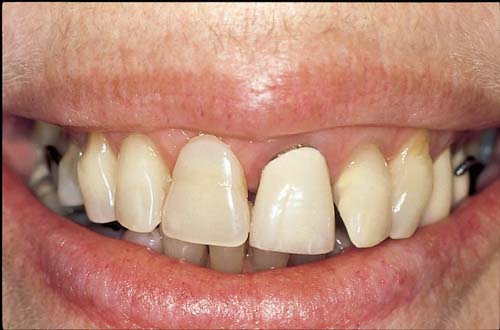 |
| Figure 2: The maxillary incisal curve and gingival plane both fail to harmonize with the lower lip curve, and the midline is not oriented vertically in the face, all contributing to an unattractive smile. |
 |
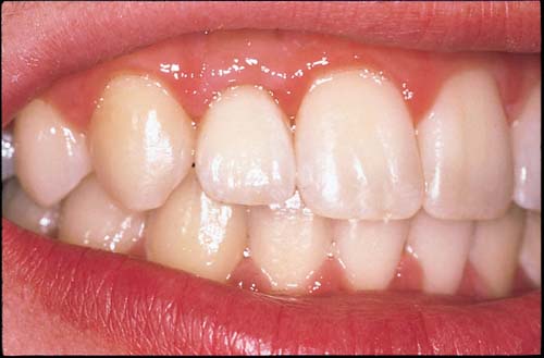
| Figure 3: Correct positioning of the maxillary central incisal edges is critical to the esthetic and functional potential of any dentition. In an intact Class I occlusion, the maxillary central incisal edges normally follow the same plane as the maxillary posterior buccal cusp tips. |
 |
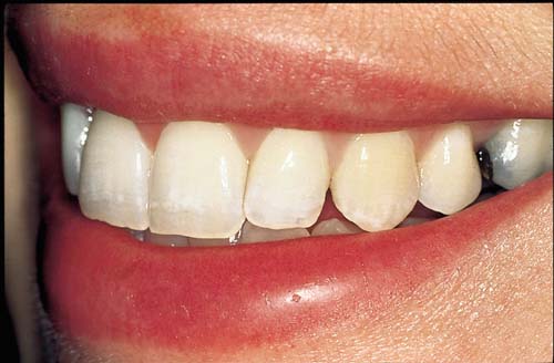
| Figure 4: Class II malocclusions commonly demonstrate overerupted maxillary central incisors, excessive incisal display and an exaggerated smile curve. |
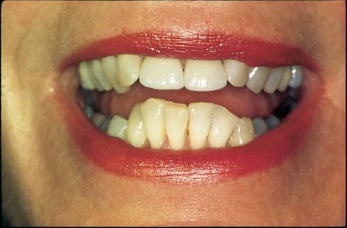 |
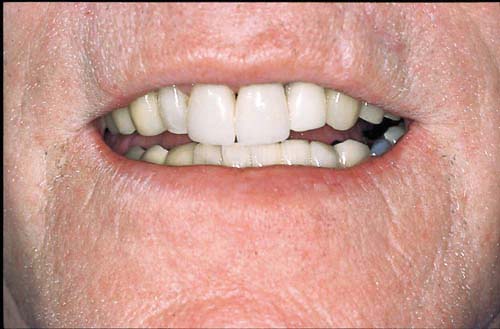
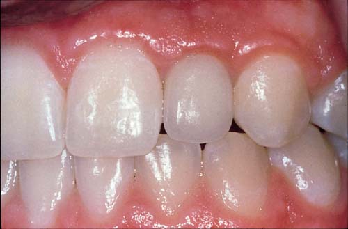
| Figure 5: Mandibular anterior overeruption resulting from a Class II malocclusion often compromises both the esthetic and the functional potential of our patients. |
 |
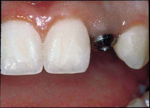
| Figure 6: This maxillary lateral incisor implant crown has been contoured interproximally to mimic natural tooth root anatomy, which controls embrasure shape and volume, thereby maintaining papilla height. |
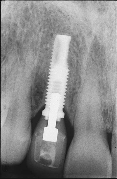 |
| Figure 7: The same patient exhibits favourable interseptal bone levels adjacent to the implant — a key factor in maintaining papilla height. The orthodontist made this possible by creating enough space between neighbouring teeth to allow for healthy interproximal bone and soft tissue following implant placement. |
 |
| Figure 8: Orthodontic distal movement of the canine creates abundant hard and soft tissue volume for implant replacement of a congenitally missing lateral incisor. |
 |
| Figure 9: Following placement of the implant crown, the same patient enjoys anatomic tissue contours resulting from initial orthodontic creation of an optimum implant site. |