|
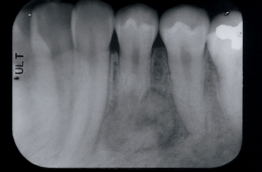
| Figure 1: Periapical radiograph of cementoblastoma associated with the left mandibular first premolar. |
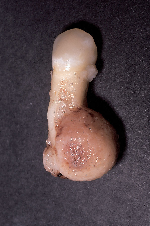 |
| Figure 2a: Buccal view of gross specimen of cementoblastoma, which is fused to the partly resorbed root of the premolar. |
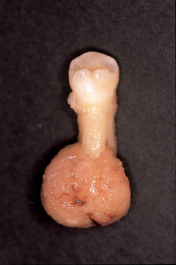 |
| Figure 2b: Lingual view of cementoblastoma. |
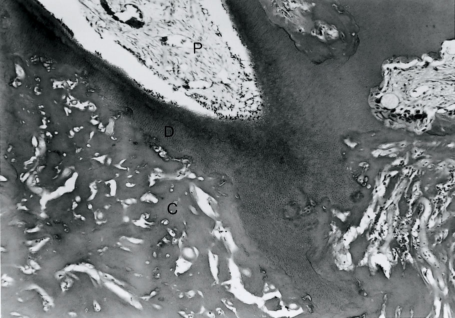 |
| Figure 3a: Resorption of the tooth root by cementoblastoma. P, vital pulp; D, dentin, which is partly resorbed and fused to cementoblastoma, C. (Hematoxylin and eosin stain of decalcified section; original magnification x 25.) |
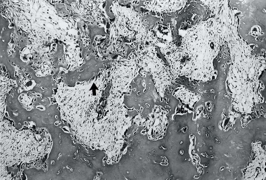 |
| Figure 3b: Moderately cellular and vascular stroma with islands and trabeculae of cementum lined by cementoblasts (arrow) and scattered cementoclasts. (Hematoxylin and eosin stain of decalcified section; original magnification x 25.) |