|
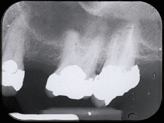
| Figure 1: Preoperative radiograph of left maxillary first molar. |
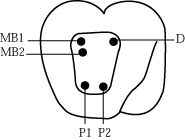 |
| Figure 2: Diagrammatic representation of the location of the orifices of the 5 canals in the maxillary left molar. |
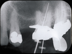 |
| Figure 3: K-type files present in the 2 palatal canals. |
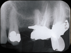 |
| Figure 4: Immediate post-treatment radiograph displaying the unique palatal morphology. |