|
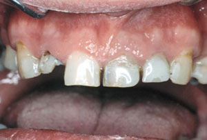
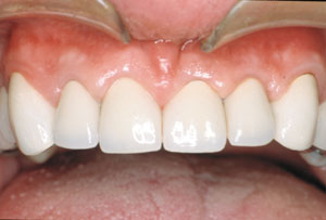
| Figure 1: Close-up view of the anterior maxillary teeth before the surgery. |
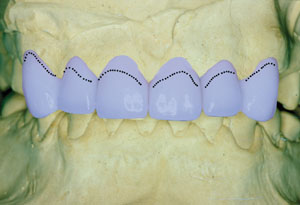 |
| Figure 2: Wax-up of the maxillary teeth. The dotted line represents the preoperative gingival margin. |
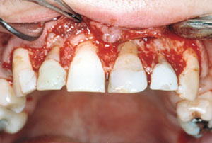 |
| Figure 3: Osseous reduction. |
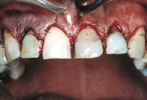 |
| Figure 4: Apically repositioned flap and sutures. |
 |
| Figure 5: Final insertion of the PFM crowns 10 months after surgery. |