Figure 1

Russel1.jpg (53149 bytes)
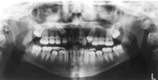
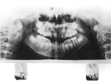
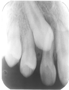
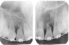
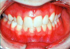
Figure 1: Clinical examination revealed asymmetry between right and left cuspids; the 53 was overretained and not mobile whereas the 63 had exfoliated naturally allowing the 23 to erupt into the arch. The 13 was palatally impacted and required surgical exposure and orthodontic alignment.


Figure 2

Russel2.jpg (18544 bytes)
Figure 2: Significant asymmetry of the eruptive stages of maxillary permanent cuspids may be a sign of potential impaction. The 13 was palatally impacted and required surgical exposure and orthodontic alignment.


Figure 3

russel3.jpg (21101 bytes)
Figure 3: The degree of asymmetry of the eruption of the 13 and 23 and the overlap of the 13 with the 12 were signs of impaction.Panoramic and periapical radiographs were used to locate the 13 on the palate.


Figure 4

Russel4.jpg (32832 bytes)
Figure 4: Significant overlap of the maxillary cuspid and the lateral incisor makes recovery of the impacted canine following extraction of the primary cuspid less likely.


Figure 5

russel5.jpg (21270 bytes)
Figure 5: These periapical radiographs show severe lateral root resorption that resulted from palatally impacted cuspids that resorbed the adjacent roots.