 |
| Figure 1: Diagram depicting the difference between incisal and marginal rests. |
|
| Figure 1: Diagram depicting the difference between incisal and marginal rests. |
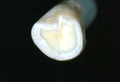 |
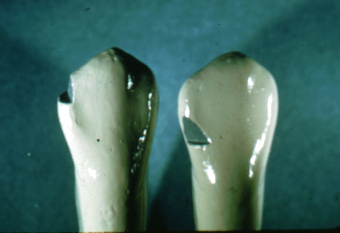
| Figure 2: Cross-sectional view of a canine crown. The left side has been sectioned in the middle third, incisogingivally. The right side has been sectioned in the gingival third. The enamel thickness in the middle third on the marginal ridge is evident. |
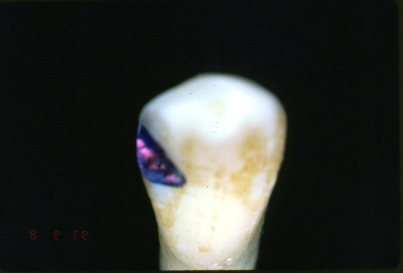 |
| Figure 3: The marginal rest in a natural canine crown. The rest seat is shaded. |