• John Carpendale, B.Dent.Sc., MSD, Dip. Pros. •
© J Can Dent Assoc 1999; 65:576-8
[Root Caries |Recurrent Caries|Principles of Mangement |Case Report|Conculsion |References]
As we approach the millennium, much is spoken about the decline in dental caries among the younger population and the long-term effects that this decline may have on the profession. Yet there is a tendency to ignore our aging population with its often extensively restored dentitions. The management of restoration failure will become an ever-increasing challenge to the profession. In many instances, recurrent caries will result in the loss of single or multiple dental units. The detection of recurrent caries adjacent to radiopaque restorations remains a challenge to our profession.
The patient with a failed restored dentition presents for one or more of the following reasons1:
• pain, usually affecting the mouth, face or temporomandibular joint
• inability to function, which can be total (i.e., the dentition is unusable) or localized (such as painful teeth, highly mobile teeth or speech problems)
• dissatisfaction with esthetics
• broken teeth or restorations
• inflammatory swelling
• bad taste
• bad breath (halitosis), having been informed by another person
• bleeding gums
• anxiety, which can be either primarily of dental origin (e.g., lost teeth or restorations) or of psychogenic origin but aggravated by dental treatment.
Additionally, the patient could be symptom-free, but have been referred for a restorative problem.
Root caries is an emerging challenge to the dental professions because of the growing number of increasingly aging adults who have retained many or all of their teeth.2 Risk factors for developing root caries point to both intraoral and environmental factors, making the management of root caries complex and multidisciplinary.3 Caries may be present beneath restorations, at the margins of restorations or on the roots.
Root surface caries is strongly associated with gingival recession and periodontal pockets. In a study of 196 dentates with a mean age of 79.3, root caries was found in 52% of the men and 35% of the women.4
Surprisingly, there was a statistically significant negative correlation between root caries incidence impact and the number of daily medications taken.
Schwartz and others5 and Randow and others6 both reported caries to be the most frequent cause of failure (36%; 18.3%) of existing restorations. In 1993, Glantz and others7 reported that of 77 bridges reviewed at 15 years, 32.5% required removal. Of these, 9.6% were removed because of untreatable caries of the abutments. In a second report, Glantz and Nilner8 noted that the incidence of caries was not related to the age of the patient but, rather, to the time that the bridge had functioned.
Often, endless discussion with the patient revolves around whether the caries is due to poorly fitting crowns. From the management point of view, however, the important aspect is that the patient has demonstrated caries susceptibility. Not all patients with poorly fitted crowns develop caries nor do well-fitting crowns provide caries immunity in the remaining tooth structure. Nonetheless, Karlsson9 reported a higher incidence of caries around crowns with poor margins compared to those with good margins.
An assessment of disease susceptibility and control is essential before formulating a definitive plan of treatment. Unfortunately, as is clearly pointed out by Bibby10 and Anderson and others,11 the diagnostic predictors for dental caries are poor.
After restoration failure caused by recurrent caries, there is often a need for restoration replacement. Careful clinical procedures and awareness of how the initial problem occurred are essential to reducing the likelihood of caries recurrence. One cannot ignore the basic principles of preparation design, careful and accurate impression procedures and their role in the production of an accurately fitting restoration.
The use of fluoride in multiple measures has a significant impact on the prevention of dental caries. These measures include public water fluoridation, professional fluoride treatments in the dental office and the home use of effective fluoridated dentifrices, with the use of fluoride rinses and gels as adjuncts when needed. In many clinical situations, professional judgment is required to identify the most appropriate treatment measures to address the needs of individual patients. Prevention based on a composite of risk factors is the most desirable approach for management.
Patients who have developed caries can be treated with remineralization strategies, recontouring techniques, intracoronal restorations of a variety of established and recently introduced materials, or extracoronal restorations.12
Often, despite careful and thorough maintenance, there can be a restoration failure. The loss of single or multiple dental units often makes restoration replacement with a fixed alternative impossible. A difficult but unavoidable transition occurs from a fixed to a removable prosthodontic appliance. The case report below illustrates the general principle of such a transition.
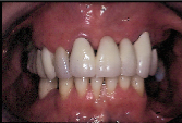
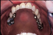
The patient, a 59-year-old woman, presented for consultation about the status of her maxillary residual dentition (Fig. 1). A suspicion of recurrent caries, coupled with her anxiety of being forced to wear a removable appliance (Fig. 2), prompted the visit.
 |
 |
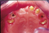 |
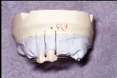
| Figure 1: Frontal view of patient’s dentition upon presentation. | Figure 2: Palatal view of patient’s dentition upon presentation. | Figure 3: Palatal view of patient’s dentition following removal of fixed bridge. |
Clinically, caries was detectable at the palatal margins of the restored maxillary dentition. The patient was partially dentate in both the maxilla and the mandible. The maxillary arch was restored with a fixed bridge reconstruction. The mandibular edentulous areas were restored with a cast framework removable partial denture.
To respond to the patient’s anxiety, it was decided to explore the status of the underlying teeth undetectable to radiographic interpretation. It was planned that the patient would leave the office with a provisional fixed partial denture. Removal of the existing full-coverage restorations revealed that the incidence of caries was more extensive than originally believed. Complete caries removal was not effected at this time (Fig. 3), as it would have eliminated the available tooth structure and prevented the opportunity for placing a provisional fixed partial denture. In consultation with the patient, it was decided to direct treatment toward the salvage of certain teeth as overdenture supports and toward the provision of an immediate complete upper denture.
Using a 2-mm-wide reinforcement ribbon (Connect, Kerr Manufacturing Co., Orange, CA), a provisional fixed partial denture was fabricated (Integrity, Dentsply/Caulk, Milford, DE) and placed using a provisional luting cement (Temp-Bond, Kerr Manufacturing Co., Orange, CA).
It was clear that oral hygiene compliance needed reinforcement. This reinforcement included reminders of basic home care procedures as well as the provision of a thermoplastic template to assist stannous fluoride home application. Diet modification was suggested.
The patient was provided with her treatment options and prognosis, with the definitive goal being the provision of endosseous implants to support a fixed maxillary reconstruction. For the patient, the cost of such long-term treatment goals was prohibitive; therefore, it was decided to retain as many teeth as possible as overdenture abutments to maintain the integrity of the alveolar process. This would have implications for denture design.
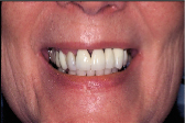 |
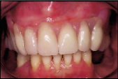
| Figure 4: Frontal view showing patient’s high smile line. |
The patient’s very high smile line (Fig. 4) in conjunction with a poor alveolar ridge height posteriorly meant that the retention of anterior abutments would imply the absence of a labial flange to the complete denture. Such a deficiency in denture design has implications for retention and denture stability. The reliance on certain remaining teeth to facilitate overdenture support and retention would become increasingly important.
Primary impressions were made with an alginate impression material. At the next appointment, on removal of the provisional bridge, it was decided to retain teeth 11, 12 and 23 as strategic teeth to act as overdenture abutments. The overdenture abutment design for teeth 11 and 12 was chosen as a splinted metal ceramic alloy with Hader Clip (Attachments International, San Mateo, CA) retention on the mesial of tooth 11 and distal of tooth 12. It was decided to use a directly placed pressed stud precision attachment (Zaag Root, Zest Attachment, Escondido, CA) for overdenture retention at tooth 23. Teeth 13 and 24 were kept as overdenture abutments but were sealed with a composite restorative material.
At a subsequent appointment, on removal of the provisional fixed bridge, a custom acrylic impression tray was border-moulded using a heavy-viscosity polyvinyl siloxane impression material (Reprosil, Dentsply/Caulk, Milford, DE), followed by a light-viscosity material to facilitate the recording of the prepared abutment teeth at teeth 11 and 12. The maxillary master cast was subsequently mounted on a semi-adjustable articulator at the patient’s current vertical dimension. The proposed denture teeth were selected with patient assistance. The teeth were set in wax and demonstrated to the patient for acceptance. A silicon mould was made of the approved tooth arrangement, the confines of which facilitated the design of the restoration on the overdenture abutments at teeth 11 and 12. A metal ceramic alloy was chosen, as it facilitated the placement of opacious porcelain to the coping (Figs. 5). This would ensure that the grey metal undertone would not affect the esthetics of the anterior denture teeth (Fig. 6).
 |
 |
 |
 |
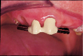
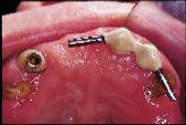
| Figure 5: Frontal view of overdenture abutment for 11 and 12 on master model. | Figure 6: Frontal view of overdenture abutment for 11 and 12 in situ. | Figure 7: Palatal view showing stud attachment in 23 and abutment in 11 and 12. | Figure 8: Frontal view of patient with complete maxillary denture. |
On the day of denture delivery, selected teeth were removed and teeth 13 and 24 were reduced to gingival level. They were subsequently sealed with composite restorative material (Z100, 3M, St. Paul, MN). Tooth 23 was reduced to gingival level and the root face prepared for the delivery of a selected stud attachment (Zaag Root) (Fig. 7). The attachment was luted in place using a resin-reinforced glass ionomer luting cement (Fuji Plus, GC America, Chicago, IL). The retention coping was also placed at teeth 11 and 12 using a resin-reinforced glass ionomer luting cement (Fuji Plus). The finished immediate complete maxillary denture was then delivered after occlusal adjustment refinement and elimination of obvious pressure areas (Fig. 8). Subsequent appointments were necessary to allow an assessment of patient adaptation and correction of denture anomalies.
A patient’s transition from having the security and comfort of a fixed appliance to having a removable appliance presents a significant challenge to the patient-dentist relationship. The emotive issues of age and personal image demand a caring and thoughtful approach to patient management. If the long-term treatment goal is the use of a fixed implant-supported prosthesis, then the adjustment to the removable appliance as a transitional appliance is acceptable. If this goal is impossible because of practical treatment planning or simple lack of finances, there must be a greater emphasis on supportive management to ensure optimal success.
A careful explanation of treatment protocols from the first visit is imperative to better patient understanding. Failure to explain adequately may result in the patient having a negative self-image, thwarting the best efforts of the practitioner to ensure success.a
Dr. Carpendale maintains a private prosthodontic practice in Vancouver, B.C.
Reprint requests to: Dr. John Carpendale, 704-2525, Willow St., Vancouver, BC V5Z 3N8
The author has no declared financial interest in any company manufacturing the types of products mentioned in this article.
1.Wise MD. Failure in the restored dentition: Management and treatment. London: Quintessence; 1995.
2. Shay K. Root caries in the older patient: significance, prevention, and treatment. Dent Clin North Am 1997; 41:763-93.
3. Powell LV, Leroux BG, Persson RE, Kiyak HA. Factors associated with caries incidence in an elderly population. Community Dent Oral Epidemiol 1998; 26:170-6.
4. Narhi TO, Vehkalahti MM, Siukosaari P, Ainamo A. Salivary findings, daily medication and root caries in old elderly. Caries Res 1998; 32:52-9.
5. Schwartz N, Whitsett L, Berry R, Stewart J. Unserviceable crowns and fixed partial dentures: life-span and causes of loss of serviceability. JADA 1970; 81:1395-1401.
6. Randow K, Glantz PO, Zoger B. Technical failures and some related clinical complications in extensive fixed prosthodontics. An epidemiological study of long-term clinical quality. Acta Odontol Scand 1986; 44:241-55.
7. Glantz PO, Nilner K, Jendresen MD, Sundberg H. Quality of fixed prosthodontics after 15 years. Acta Odontol Scand 1993; 51:247-52.
8. Glantz PO, Nilner K. Patient age and long-term survival of fixed prosthodontics. Geriodontology 1993; 10:33-9.
9. Karlsson S. A clinical evaluation of fixed bridges, 10 years following insertion. J Oral Rehabil 1986; 13:423-32.
10. Bibby BG. Methods of Caries Prediction. Information Retrieval Inc. Washington DC and London. 1977.
11. Anderson MH, Bales DJ, Omnell KA. Modern management of dental caries: the cutting edge is not the dental bur. JADA 1993; 124:36-44.
12. Stookey GK. Caries prevention. J Dent Educ 1998; 62:803-11.