

![]()
![]()
The Use Of Cyanoacrylates In Periodontal Therapy
Jim Grisdale, BA, DDS, Dip. Prosth., Dip. Perio., MRCD(C)
ABSTRACT
N-butyl cyanoacrylate is an effective tissue adhesive which is hemostatic and
bacteriostatic. It can be considered an alternative to conventional sutures in soft-tissue
surgery. The author presents two cases demonstrating the use of the material. Case One
shows its use in free gingival graft surgery. Case Two shows its use post-biopsy.
MeSH Key Words:cyanoacrylates/therapeutic use; periodontal dressings; tissue
adhesives.
© J Can Dent Assoc 1998; 64:623-3
This article has been peer reviewed.
[
Introduction | Case One | Case Two | Acknowledgements]
Extensive research is available from the early 1960s on dental uses of cyanoacrylate materials. Several forms of cyanoacrylate have been studied for possible use in humans. Alpha and ethyl forms were found incompatible for use in humans. Particular interest has focused on the use of n-butyl cyanoacrylate, which is biocompatible in humans. This material is an effective tissue adhesive which displays hemostatic properties and a bacteriostatic action.
N-butyl cyanoacrylate has several advantages over conventional suture materials in soft-tissue surgery: it saves time, is hemostatic and bacteriostatic, and doesn’t need to be removed during post-operative follow-up. Patients must wear eye protection when cyanoacrylates are being applied, as they may cause irreversible retinal damage.
In my practice, I use cyanoacrylate to facilitate the free gingival graft surgical procedure (Fig. 1(a)). Following preparation of the recipient site, the donor tissue is obtained from the palate. Cyanoacrylate is then applied as a dressing over the donor site. The material is useful here because of its hemostatic and bacteriostatic properties and because it acts as a protective barrier during the healing phase (Fig. 1(b)).
Following accurate positioning of the donor tissue in the recipient site, I apply digital pressure for approximately four minutes. Using a special applicator, I then apply cyanoacrylate at the coronal border of the recipient site/donor tissue interface, to aid fixation (Fig. 1(c)). The distinctive color of the tissue adhesive, facilitates this part of the procedure. The appropriate applicator allows easy, controlled placement of the material. Care should be taken to prevent the cyanoacrylate from flowing between the juxtaposed tissues. Excess material may be removed by “blotting” with a cotton-tip applicator. One or two layers of cyanoacrylate should suffice to create a bond between the tissues.
A further periodontal dressing may be placed over the recipient site at the clinician’s discretion. Post-operative care instructions are given to the patient and appropriate follow-up appointments are arranged (Fig. 1(d)). The cyanoacrylate material I prefer to use for this technique is Periacryl (Blacklock Medical Products, Delta, B.C.). I prefer it because of its distinctive color, consistency of flow, ease of application and the consistent post-operative results it produces. It is also relatively inexpensive.
Practitioners may have the occasion to perform a soft tissue biopsy and submit the sample for histologic examination and diagnosis (Fig. 2(a)). The use of sutures for wound closure may be very difficult or impractical in certain situations. Periodontal dressings may be difficult to apply and retain. Bleeding may also be difficult to control. In my practice, following the soft-tissue biopsy, I apply a thin layer of cyanoacrylate to the biopsy site. The material is effective as a surgical dressing during the early healing phase. (Fig. 2(b)). Appropriate post-operative care instructions are given and the patient is asked to return for follow-up (Fig. 2(c)).
I have found cyanoacrylate to be useful in several other periodontal surgery applications. These include post-gingivectomy, gingivoplasty and tooth extraction; securing apically positioned flaps; and following ridge preservation procedures using osseous graft materials.
Dr. Grisdale is in private practice and is affiliated with the continuing dental eduction department at UBC.
Reprint requests to: Dr. Jim Grisdale, 805-805 West Broadway, Vancouver, BC V5Z 1K1.
The author has no declared financial interest in any company manufacturing the types of products mentioned in this article.
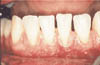
Fig 1a: Preoperative view of graft recipient site.
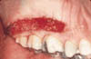
Fig 1b: Donor site with cyanoacrylate dressing in-situ.
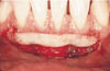
Fig 1c: Cyanoacrylate anchoring the donor tissue in place.
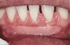
Fig 1d: Postoperative view of the healed recipient site.
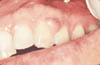
Fig 2a: Preoperative view of biopsy site.
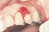
Fig 2b: Cyanoacrylate applied to biopsy site.
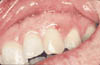
Fig 2c: Healed biopsy site at follow-up.