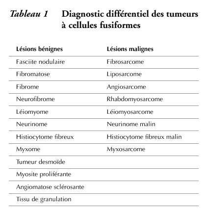
|
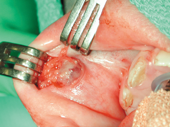
| Illustration 1 : Exposition d’une lésion intra-buccale d’évolution rapide chez une fillette de 9 ans. |
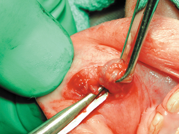 |
| llustration 2 : Ablation de la lésion. |
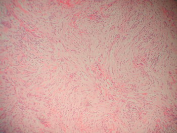 |
| Illustration 3 : Examen histopathologique par coloration à l’hématoxyline-éosine (H et E), montrant la disposition storiforme (grossissement initial x 100). |
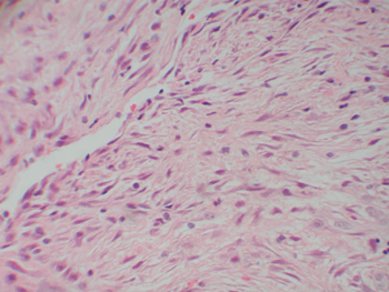 |
| Illustration 4 : Examen histopathologique par coloration à H et E montrant la présence de cellules fusiformes (grossissement initial x 400). |