40
|
Volume2 Issue2
S
upporting
Y
our
P
ractice
Thrombocytopenicpurpura
•
Decreasedplatelet production (e.g.,myelofibrosis,
leukemia,myelodysplastic syndromes, chemotherapy,
radiotherapy, skeletalmetastasis)
• Increasedplatelet destruction (e.g., immune
thrombocytopenicpurpura)
• Platelet sequestration (e.g., splenomegaly)
Disordersof coagulation
•
Inherited (e.g., hemophiliaA andB)
• Acquired (e.g., liver disease, vitaminKdeficiency,
medication [warfarin, heparin])
Treatment
Common Initial Treatment
1.
Bleedingcanusuallybecontrolledby localmeasures:
• Pressurewithmoistenedgauze
• Absorbablegelatin, absorbable collagen,microfibrillar
collagenor oxidized regenerated cellulose (toprovide
a scaffold for platelets to adhere)
• Thrombin (to convert fibrinogen tofibrin)
• Epsilon-aminocaproic acidor tranexamic acidoral
rinses (to reducefibrinolytic activity)
• Soft diet and avoiding factors thatmayprovoke
bleeding, such as strenuous activities, traumatic
brushing, flossing, and rinsing
• If pre-existingdentalmodels are available, a vacuum-
formed splint (+/− linedwith thrombinpowder) can
be fabricated andused to apply additional pressure
andprotection
2.
Screening serologic studies shouldbeperformed.
• Completeblood count (CBC)withplatelet count
• International normalized ratio (INR):measures the
factors of theextrinsic and common coagulation
pathways
• Partial thromboplastin time (PTT):measures the
factors of the intrinsic and common coagulation
pathways
• Thrombin time (TT): tests the abilityof fibrinogen to
form an initial clot
• Platelet function analyzer (PFA-100) or Ivybleeding
time (BT): screens for functional platelet disorders
3.
If thebleedingcannot becontrolled, assessmentwitha
physicianand systemicmeasures arenecessary.
4.
Definitivemedicalmanagement dependson thenature
of theunderlyingdisorder. Principal agents for systemic
management includeplatelet infusion, fresh frozenplas-
ma, factor concentrates, cryoprecipitate, desmopressin
(DDAVP) andantifibrinolytic therapy.
2
. Inspect thevisible skinandperforman intraoral
examination.
3.
Assess for other potential causesof oral hemorrhage:
• Mucocutaneousdisorders (e.g., desquamative
gingivitis [erosive lichenplanus, pemphigus vulgaris,
mucousmembranepemphigoid anderythema
multiforme])
• Necrotizingulcerativegingivitis
• Drug-inducedgingival hyperplasia
• Gingivitis of a local or endocrine (puberty, pregnancy)
cause
• Periodontitis
Hemorrhage that isevokedwithminimal provocationor
spontaneously, especially ifprolongedanddifficult tocontrol,
shouldalert theclinician toanunderlyingbleedingdisorder.
Diagnosis
•
Ahematologistwill performa focusedhistory, physical
examination, and laboratory studies thatmay include
specificcoagulation factor assays,mixing studies and
platelet aggregation tests.
•
If leukemia is suspected, diagnosis is confirmedby
peripheral blood smear andbonemarrowbiopsy for
cytology, immunophenotyping, andmolecular/
cytogenetic studies.
Differentialdiagnosis
•
Local pathologies (see the Investigation section)
•
Systemicdisorders:
Nonthrombocytopenicpurpura
• Vascular disorders (e.g., scurvy, Ehlers-Danlos syndrome,
hereditaryhemorrhagic telangiectasia)
• Disorders of platelet function (e.g., inheriteddisorders
[Bernard-Soulier syndrome, vonWillebranddisease],
medications [ASA, NSAIDs], alcoholism, uremia)
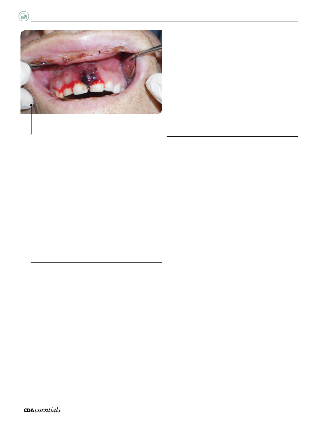
Fig.1:
Leukemicgingivitiswithprolongedgingival hemorrhage
followingminor provocation.


