 |
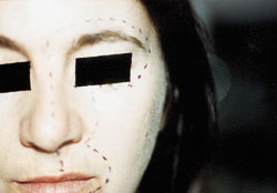
| Figure 1: Numbness in left supra-orbital region and area innervated by the second and third divisions of the left trigeminal nerve (affected zones outlined by dots). |
|
| Figure 1: Numbness in left supra-orbital region and area innervated by the second and third divisions of the left trigeminal nerve (affected zones outlined by dots). |
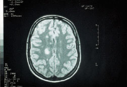 |
| Figure 2: Magnetic resonance image confirming the presence of demyelinating lesions in the cerebral hemispheres (dense white areas). |
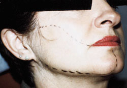 |
| Figure 3: Numbness of the lower half of the right side of the face (affected zone outlined by dots). |
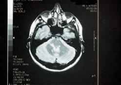 |
| Figure 4: Magnetic resonance image confirming the presence of lesions in the brain stem and the median cerebellar peduncles. |