

![]()
![]()
Porcelain Veneers:a Challenging Case
Daniel J.J. Fortin, DMD,MS
ABSTRACT
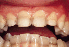
A patient in his early 20s with teeth badly discoloured by tetracycline was seeking
treatment to improve his esthetics. Because retreatment and cost were important
considerations, porcelain veneers were the treatment of choice. The challenge in this case
was to mask the underlying tetracycline stain before the final cementation and thus gain
more control over the final shade of the veneers.
MeSH Key Words: case report; dental porcelain; dental veneers; tooth
discoloration/therapy.
© J Can Dent Assoc 1999; 65:110-2
[The Challenge| Possible Solutions | Porcelain Fused to Metal Crown | Porcelain Veneers |Reference]
Teeth may be discoloured as a result of tetracycline intake during a prophylactic or therapeutic regimen in the pregnant female or in the infant.
Tetracycline and similar antibiotics have a selective affinity for deposition in bone and tooth substance.1
The portion of the tooth stained by tetracycline is determined by the stage of tooth development at the time of drug administration. The discoloration itself depends upon the dosage, the length of time over which administration occurred and the type of tetracycline. The teeth affected by tetracycline appear to have a yellowish or brownish-grey discoloration, which is most pronounced at the time of eruption of the teeth. This discoloration gradually becomes more brownish after exposure to light.2
Success in bleaching varies depending on the individual and the etiology of the discoloration. Bleaching vital teeth is a safe procedure, but results are variable and hard to predict, especially for teeth showing tetracycline banding as in this case.3
Typical indications for veneers include teeth that are malformed, rotated or malpositioned. Veneers may also be used to close single or multiple diastemas to change the colour of unattractive teeth, to restore abraded or eroded restorations or to cover and replace faulty restorations. Other factors such as stain etiology, occlusion and the age, health and oral hygiene of the patient are important considerations in the treatment plan.
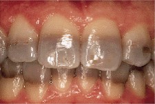
Masking tetracycline stains is one of the ultimate tests for porcelain veneers. It is difficult to mask dark underlying tooth colour and retain a natural appearance of the veneers. A very important factor in successfully covering these stains is the area of each tooth that is affected. Staining of the incisal third or the middle third of the teeth is relatively easy to cover. Staining of the gingival third is a difficult situation for veneers. The challenge in this case was to mask the underlying tetracycline stain before the final cementation, which enabled more control over the final shade of the veneers.
Some operators prefer to etch the tooth and apply the veneer directly over the entire untouched facial surface, thereby not removing any enamel. Several problems exist with such a method. The reversibility of these veneers may seem desirable, but the esthetic results and physiological contours are not always optimal. In fact, restorations are usually over-contoured and gingival inflammation may be observed. The removal of some enamel before placing a veneer is recommended to achieve ideal esthetic and physiological results. A clear finish line and specific surface reductions will facilitate laboratory fabrication and cementation.
Teeth that have been stained by tetracycline are more difficult to mask with veneers, especially when the cervical areas are badly discoloured. This complex problem requires a specific approach. The preparation should be conservative. It should allow space for a coverage of 0.5 mm to 0.75 mm of porcelain. Any area of the tooth that is visually accessible should be covered by porcelain. If the defect does not extend subgingivally, the margin of the veneer should remain above the gum line. Veneer margins will blend with the gingival enamel and impression making will be easier.
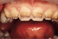
Incisal reduction is an important factor in the long-term fracture resistance of veneers.4 The incisal edge is reduced by 1.5 mm and a lingual chamfer is prepared (Figs. 2 and 3). This chamfer exposes porcelain to compression instead of shearing during the initial phase of protrusive movement, and as long as forces are against the tooth (compressed porcelain), fracture resistance is high.
 |
 |
| Fig. 2: Lateral reduction of 0.5 to 0.75 mm. | Fig. 3: Incisional edge is reduced by 1.5 mm with a lingual chamfer. |
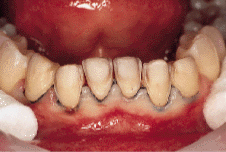
This procedure confines all the peripheral marginal areas within enamel to ensure adequate sealing by normal bonding procedures. Final preparations should be as smooth as possible to improve accuracy of impressions and laboratory procedures. When a person smiles or talks, the six maxillary anterior teeth are usually the most noticeable; however, maxillary first premolars and mandibulary incisal are also included if there is an esthetic problem.
Generally, veneers do not penetrate into dentin. But in this case it was necessary to go deeper than just enamel, because the tetracycline banding was in the middle third of the teeth and would show through any type of opaque veneer.
As seen in this case (Fig. 2), the tooth appears darker as enamel is removed and the underlying stained dentin is exposed. To optimize the esthetic outcome, the tetracycline band needs to be opacified before cementation, or the opaqueness necessary in the porcelain to camouflage the stain will result in teeth with considerably less vitality. To cover the dark portion stained by the antibiotic, Class V type preparations were cut in dentin in the stained areas (Fig. 4), thereby removing most of the dark pigmentation responsible for the coloration. Adhesion of composite resin was achieved with a newer generation bonding system that relies on the formation of a hybrid layer to seal the exposed dentin. To maximize the opacification of the stain, a light opaque stiff composite resin was used as a restoration material (Figs. 5 and 6). Most of the darker stains are not visible anymore and should not interfere with the final colour of the veneer.
 |
 |
| Fig. 4: Class V type preparations are cut in dentin in the stained area. | Fig. 5 Opacification of the stain. |
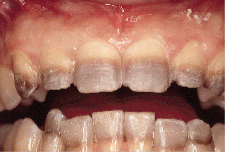
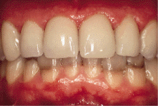
A light retraction was achieved with surgical silk. Final impressions and occlusal records were taken. The shade selection was made as if the veneers were to be on teeth of normal colour. When returned from the laboratory, the veneers were tried for fit and colour matching. No shade modification was necessary, and an untinted shade of composite cement was used for final cementation (Figs. 7 and 8).
 |
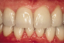 |
| Fig. 7: Immediately following surgery, after cementation of upper veneers. | Fig. 8: Upper and lower teeth immediately following surgery. |
The proper selection and manipulation of materials provide the clinician with enhanced control and predictability of esthetic restorations. In certain situations, porcelain veneers can be considered an esthetic option for severely discoloured teeth.
Dr. Fortin is director of clinical research with Dentsply/Caulk
Reprint requests to: Dr. D. Fortin, Dentsply/Caulk, L.D. Caulk Division, 38 West Clarke Ave., P.O. Box 359, Milford, Delaware 19963-0359, USA.
1. Urist MR, Ibsen HK. Chemical reactivity of mineralized tissue with oxytetracycline. Arch Pathol 1963; 76:484.
2. Shafer WG, Hine MK, Levy BM. A textbook of oral pathology, 4th ed. Philadelphia: W.B. Saunders Co.; 1983.
3. Haywood VB, Leonard RH, Dickinson GL. Efficacy of six months of nightguard vital bleaching of tetracycline-stained teeth. J Esthet Dent 1997; 9:13-9.
4. Garber DA. Porcelain laminate veneers: ten years later. Part 1: tooth preparation. J Esthet Dent 1993; 5:56-62.